
Method and system for measuring in-vivo temperature by using non-invasive technology

A technology for measuring internal body temperature and technology, applied in the medical field, it can solve the problems of poor internal temperature accuracy, lack of temperature reference system, and inability to obtain temperature information, so as to achieve the effect of eliminating trauma and pain.

Inactive Publication Date: 2020-09-08
JIESHOU JINGHUA TECH INFORMATION CONSULTING SERVICE CO LTD
View PDF0 Cites 0 Cited by
  • Summary
  • Abstract
  • Description
  • Claims
  • Application Information

AI Technical Summary

Problems solved by technology

[0002] The current temperature measurement methods are: thermocouple method, optical fiber method, infrared method, thermal paper method, liquid crystal foil method, model substance method, microwave radiation method, and nuclear magnetic resonance method. Among them, only two methods are suitable for internal anti-electromagnetic interference temperature measurement One is the nuclear magnetic resonance method, which uses magnetic resonance heat source imaging technology combined with big data to calculate through software. Although it can non-invasively obtain temperature information in the human body, due to the lack of an accurate temperature reference system in the body, according to the body surface temperature and scanning thermal imaging data The accuracy of the calculated body temperature is poor; another optical fiber method can accurately measure the temperature of certain positions in the body through invasive insertion. Although it is not subject to electromagnetic interference and can perform multi-point temperature measurement in the body, due to invasive Not only does it increase the patient's pain and infection risk, but it is also restricted by many factors and cannot reach any part of the human body (such as inside the liver, lungs, kidneys, etc.), so it is impossible to obtain temperature information of all positions inside the human body
[0003] In the field of tumor hyperthermia technology, it is particularly important to accurately measure the temperature in the patient's body. At present, all existing in vivo hyperthermia instruments cannot accurately measure the body temperature. The temperature error seriously affects the precise treatment effect of the hyperthermia equipment. Therefore, it is urgent It is necessary to provide a method for accurate and non-destructive temperature measurement in the body to solve the above problems

Method used


Image

  • Method and system for measuring in-vivo temperature by using non-invasive technology
  • Method and system for measuring in-vivo temperature by using non-invasive technology
  • Method and system for measuring in-vivo temperature by using non-invasive technology

Examples


Embodiment 1

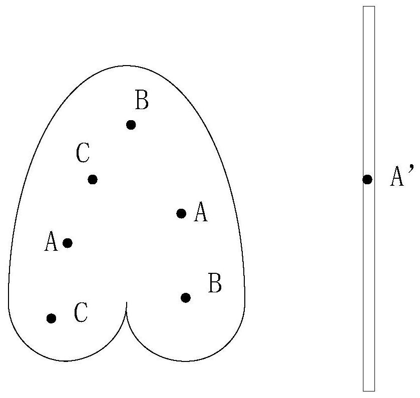
[0038] combine figure 2 It is a temperature distribution map (the figure shown is only a schematic diagram), the left side is the MRI scanning the left lung of the patient's lungs, that is, the right side is the optical fiber temperature measuring device, and the optical fiber sensor located at point A' of the optical fiber temperature measuring device The precise temperature value X=39.5°C was measured at this point. In the temperature distribution map on the left, find two points A that are identical to the thermal imaging data of point A', and the temperature value of these two points A is X=39.5°C.

[0039] According to the comparison of the thermal imaging data of each hot spot in the temperature field distribution diagram, the temperature differences between the two hot spots B and C and point A are calculated to be +0.8°C and -0.3°C respectively, then the temperature at point B is (39.5+0.8)°C or 40.3 ℃, the temperature at point C is (39.5-0.3) ℃ or 39.2 ℃.

Embodiment 2

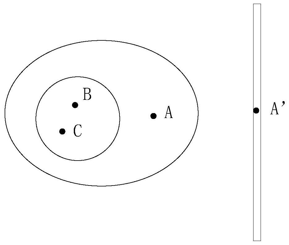
[0041] combine image 3 The temperature distribution map (shown in the figure is only a schematic diagram), the left side is the position of the patient's abdominal section scanned by the nuclear magnetic resonance instrument, and the right side is the optical fiber temperature measurement device. The optical fiber sensor at point A' of the optical fiber temperature measurement device measures the temperature Accurate temperature value X = 39.5°C. In the temperature distribution map on the left, find a point A that is the same as the thermal imaging data of point A', and the precise temperature of this point is also X=39.5°C.

[0042] According to the comparison of thermal imaging data of each hot spot in the temperature field distribution map, the temperature difference between point B and point A adjacent to A in the temperature distribution map is +0.3°C, and the precise temperature of point B is calculated to be (39.5+0.3)°C That is 39.8°C. The temperature difference bet...

Embodiment 3

[0044] same combination figure 2 The temperature field distribution diagram (shown in the figure is only a schematic diagram), the left side is the position of the patient's left lung section scanned by the nuclear magnetic resonance instrument, and the right side is the optical fiber temperature measurement device. Point A on the temperature measurement optical fiber is used as the temperature calibration point. The precise temperature value X=39.5°C measured by the optical fiber sensor at this point, and the thermal imaging data of point A obtained from the temperature field distribution map is a, assuming that there is another hot spot B in the temperature distribution map, the thermal imaging data of this point The imaging data is b. Through thermal imaging data processing, the temperature multiple of point B and calibration point A is calculated to be 1.02. With the reference of the temperature of point A at 39.5°C, the precise temperature of point B can be calculated as ...


PUM


Abstract

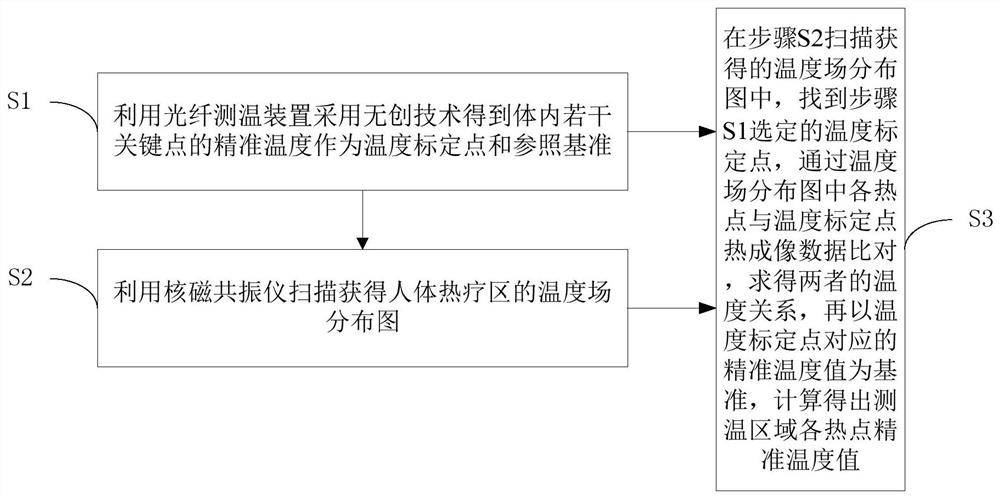
The invention discloses a method for measuring in-vivo temperature by using a non-invasive technology. The method comprises the following steps: S1, obtaining accurate temperature of a plurality of key points of a human body thermal therapy area as a temperature calibration point and a reference benchmark by using an optical fiber temperature measuring device and using the non-invasive technology;S2, performing scanning by utilizing a nuclear magnetic resonance instrument to obtain a temperature field distribution diagram of the human body thermal therapy area; and S3, finding the temperaturecalibration point selected in the step S1 in the temperature field distribution diagram obtained through scanning in the step S2, and calculating the temperature relationship between hot spots in thetemperature field distribution diagram and the thermal imaging data of the temperature calibration point through comparison, and calculating the accurate temperature values of the hot spots in the thermal therapy area by taking the accurate temperature value corresponding to the temperature calibration point as a reference. The invention also discloses a system for measuring the in-vivo temperature by using the non-invasive technology. By combining the nuclear magnetic resonance thermal imaging technology and the optical fiber temperature measurement technology, noninvasive accurate measurement of the wide-area temperature in the body is achieved, and the pain of a patient is greatly relieved.

Description

technical field [0001] The invention relates to the medical field, in particular to a method and a system for measuring body temperature using a non-invasive technique. Background technique [0002] The current temperature measurement methods are: thermocouple method, optical fiber method, infrared method, thermal paper method, liquid crystal foil method, model substance method, microwave radiation method, and nuclear magnetic resonance method. Among them, only two methods are suitable for internal anti-electromagnetic interference temperature measurement One is the nuclear magnetic resonance method, which uses magnetic resonance heat source imaging technology combined with big data to calculate through software. Although it can non-invasively obtain temperature information in the human body, due to the lack of an accurate temperature reference system in the body, according to the body surface temperature and scanning thermal imaging data The accuracy of the calculated body ...

Claims


Application Information

Patent Timeline
Patent Type & Authority Applications(China)
IPC IPC(8): A61B5/01G01K11/32G01R33/48
CPCA61B5/015G01K11/32G01R33/4804
Inventor 尚诚德
Owner JIESHOU JINGHUA TECH INFORMATION CONSULTING SERVICE CO LTD
Learn More