Breast ultrasound examination robot

An ultrasonic examination and robot technology, applied in mammography, ultrasonic/sonic/infrasonic diagnosis, ultrasonic/sonic/infrasonic Permian technology, etc., can solve problems such as low scanning efficiency, high load pressure, and shortage of doctor resources. Easy to replace and clean, reduce labor intensity

Pending Publication Date: 2020-09-25
HARBIN UNIV OF SCI & TECH
View PDF10 Cites 10 Cited by
  • Summary
  • Abstract
  • Description
  • Claims
  • Application Information

AI Technical Summary

Problems solved by technology

The sonographer repeats this non-physiological posture for diagnosis work for a long time, which will cause the body to suffer from high load pressure
[0004] To sum up, the existing ultrasonography methods have the problems of shortage of doctors' resources and low scanning efficiency.

Method used


Image

  • Breast ultrasound examination robot
  • Breast ultrasound examination robot
  • Breast ultrasound examination robot

Examples


Embodiment 1

[0028] Example 1: Automatic inspection process:

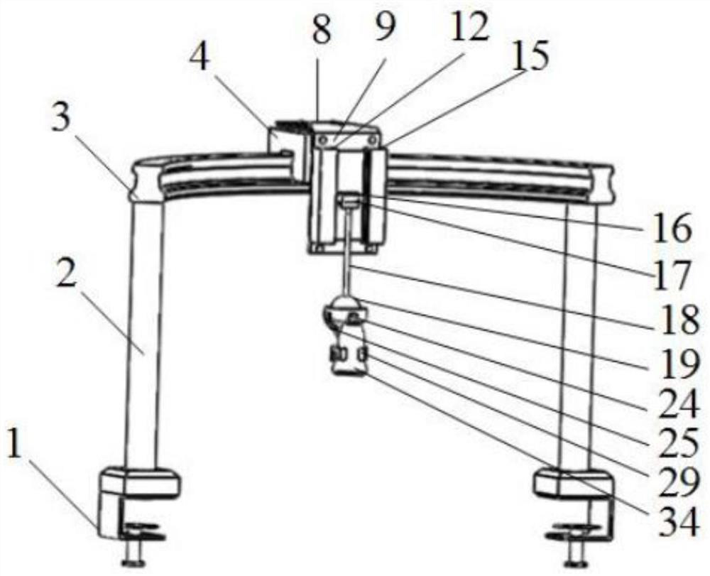
[0029]Turn on the power switch, the outer swing ring drive motor 26 controls the swing of the outer swing ring 24, the inner swing ring drive motor 27 controls the swing of the inner swing ring 25, and the outer swing ring 24 and the inner swing ring 25 act together on the ball joint 23. The swing rod 28 is stressed so that the ultrasonic probe end holder 29 clamps the ultrasonic probe 34 to perform a full range of ultrasonic scans on the patient;

Embodiment 2

[0030] Embodiment 2: Auxiliary inspection process:

[0031] Adjust the device to a suitable position, turn off the power switch, and the doctor can operate the robot to perform ultrasonic scanning on the interested part according to his own experience. The grooves on the surface are in contact with each other, and under the action of the ball joint positioning spring 22 between the ball joint positioning rod 21 and the positioning sleeve 20, the robot assists the doctor in fixing the position of the ultrasonic probe, reducing the doctor's work intensity.


PUM


Abstract

The invention discloses a breast ultrasound examination robot. The breast ultrasound examination robot comprises the components: an ultrasonic probe position adjustment module and an ultrasonic probeposture adjustment module, wherein the ultrasonic probe position adjustment module comprises a circular arc sliding rail, a sliding block, a feeding screw, a feeding sliding table, lifting screws, lifting sliding tables, screw motors and a lifting rod; and the ultrasonic probe posture adjustment module comprises a spherical joint sleeve, a spherical joint, an inner swing ring, an outer swing ring,drive motors, a swing rod and a tail end clamping plate. According to the breast ultrasound examination robot, automation of breast ultrasound scanning can be achieved, and an ultrasonic probe can bekept hovering at an arbitrary position within the working range and does not need to be held by a doctor all the way, so that the scanning efficiency is improved and the labor intensity of the doctoris reduced.

Description

technical field [0001] The invention belongs to the field of medical devices, in particular to a breast ultrasound examination robot. Background technique [0002] Breast ultrasonography is the most characteristic for the identification of cystic masses or solid masses in the breast, and is of great significance for the screening of early breast cancer. Therefore, breast ultrasonography is widely used in clinical practice. [0003] Ultrasound examination is a repetitive work with high fatigue intensity. The daily work of ultrasonography is very heavy. Doctors are prone to repetitive labor injuries when they work for a long time. During the examination, doctors need to hold the probe and place it on the part to be tested. In addition, it is necessary to operate the machine to freeze the image and adjust imaging parameters in real time according to the imaging effect, such as imaging depth, gain, mode, etc., which often makes the doctor's body appear uncomfortable. Sonographe...

Claims


Application Information

Patent Timeline
Patent Type & Authority Applications(China)
IPC IPC(8): A61B8/08A61G7/05
CPCA61B8/0825A61B8/4209A61G7/05
Inventor 张永德赵梓汐赵佳伟高英博
Owner HARBIN UNIV OF SCI & TECH
Why Eureka
Try Eureka