Eye fundus image blood vessel segmentation method of semantic and multi-scale fusion network

A multi-scale fusion, fundus image technology, applied in the field of medical image processing and computer vision, can solve problems such as large changes in blood vessel scale, and achieve the effect of simple program, easy construction and high accuracy

Pending Publication Date: 2020-10-02
DALIAN UNIV OF TECH
View PDF0 Cites 4 Cited by
  • Summary
  • Abstract
  • Description
  • Claims
  • Application Information

AI Technical Summary

Problems solved by technology

The semantic fusion module improves the segmentation accuracy of capillaries by better fusing high-dimensional semantic information, and the multi-scale fusion module uses the fusion of multi-scale information to solve the problem of large changes in blood vessel scale

Method used


Image

  • Eye fundus image blood vessel segmentation method of semantic and multi-scale fusion network
  • Eye fundus image blood vessel segmentation method of semantic and multi-scale fusion network
  • Eye fundus image blood vessel segmentation method of semantic and multi-scale fusion network

Examples


Embodiment Construction

[0030] The present invention proposes a method for segmenting blood vessels in fundus images based on semantic and multi-scale fusion networks. The details are as follows in conjunction with the accompanying drawings and embodiments:

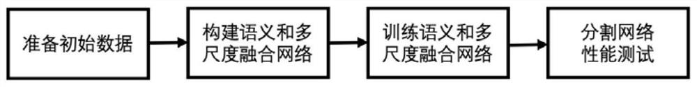
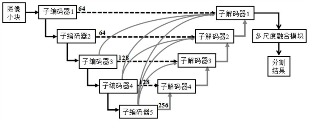
[0031] The present invention builds a semantic and multi-scale fusion network, uses fundus images for training, and achieves a high segmentation accuracy rate in the test. The specific implementation process is as follows figure 1 As shown, the method comprises the following steps;

[0032] 1) Prepare the initial data: process the retinal fundus data to generate small fundus image patches for training and testing and image patches corresponding to the blood vessel segmentation labels of the fundus images.

[0033] 1-1) Two public datasets of fundus images are used, namely CHASE_DB1 (Fraz M M, Remagnino P, Hoppe A, et al. An ensemble classification-based approach applied to retinal blood vessel segmentation[J]. IEEE Transactions on Biomedical Eng...


PUM


Abstract

The invention discloses an eye fundus image blood vessel segmentation method of a semantic and multi-scale fusion network, and belongs to the field of medical image processing and computer vision. According to the method, a semantic fusion module and a multi-scale fusion module are designed, and the semantic fusion module and the multi-scale fusion module are utilized to construct a semantic and multi-scale fusion network with a unique structure for segmenting fundus vessels of retinal images. The semantic fusion module improves the segmentation precision of capillaries by better fusing high-dimensional semantic information, and the multi-scale fusion module solves the problem that the blood vessel scale change is large through fusion of multi-scale information. Experiments prove that themethod can effectively improve the retinal fundus vessel segmentation precision. In addition, the network provided by the invention is clear in structure, easy to construct and easy to implement.

Description

technical field [0001] The invention belongs to the field of medical image processing and computer vision, and relates to segmenting blood vessels in fundus images by using a deep learning neural network framework, in particular to a method for segmenting blood vessels in fundus images with semantic and multi-scale fusion networks. Background technique [0002] Retinal diseases, such as diabetic retinopathy and glaucoma, are the leading causes of blindness and a major public health concern worldwide. Due to lifestyle changes, population aging and other risk factors, the number of patients with retinal diseases is gradually increasing. This has driven many research efforts to develop computer-aided diagnosis (CAD) systems to automatically diagnose retinopathy. Segmentation of retinal vessels is a fundamental step in building such CAD systems and is critical for accurate quantification of retinal diseases on fundus images. Although substantial work has been devoted to the se...

Claims


Application Information

Patent Timeline
Patent Type & Authority Applications(China)
IPC IPC(8): G06T7/11G06T7/194
CPCG06T2207/20081G06T2207/20084G06T2207/30041G06T2207/30096G06T2207/30101G06T7/11G06T7/194
Inventor 徐睿叶昕辰姜桂良刘恬恬
Owner DALIAN UNIV OF TECH
Why Eureka
Try Eureka