Anterior segment sectional image feature extraction method based on machine vision

A tomographic image and feature extraction technology, applied in image enhancement, image analysis, image data processing and other directions, can solve problems such as inability to cooperate, eyes unable to focus, and measurement accuracy decline, to enhance image contrast and illumination equalization, The effect of reducing discomfort, improving fit and comfort

Pending Publication Date: 2020-10-30
THE EYE HOSPITAL OF WENZHOU MEDICAL UNIV
View PDF7 Cites 3 Cited by
  • Summary
  • Abstract
  • Description
  • Claims
  • Application Information

AI Technical Summary

Problems solved by technology

[0002] When using a slit lamp or related equipment to collect anterior segment tomographic images, in order to ensure image quality, a high-intensity light source is usually used to irradiate the human eye. Some people who are sensitive to light cannot even cooperate to complete the entire detection, requiring the cooperation of an eye opener, and the eyes cannot be used during the test. Focus on the point of gaze, resulting in a decrease in measurement accuracy

Method used


Image

  • Anterior segment sectional image feature extraction method based on machine vision
  • Anterior segment sectional image feature extraction method based on machine vision
  • Anterior segment sectional image feature extraction method based on machine vision

Examples


Embodiment Construction

[0031] The present invention will be described in further detail below in conjunction with the accompanying drawings.

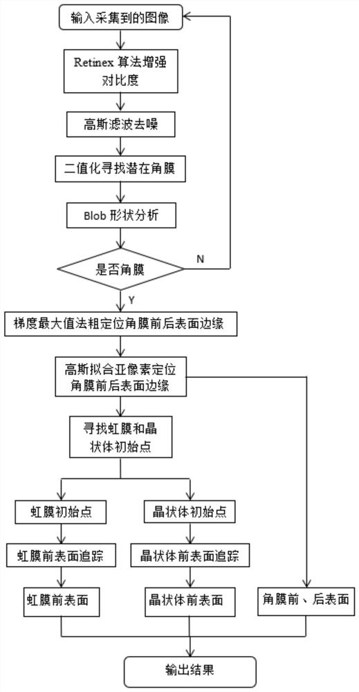
[0032] Reference attached figure 1 , a method for feature extraction of anterior segment tomographic images based on machine vision, comprising the following steps:

[0033] Step S1. Under low-illumination lighting, collect anterior segment tomographic images;

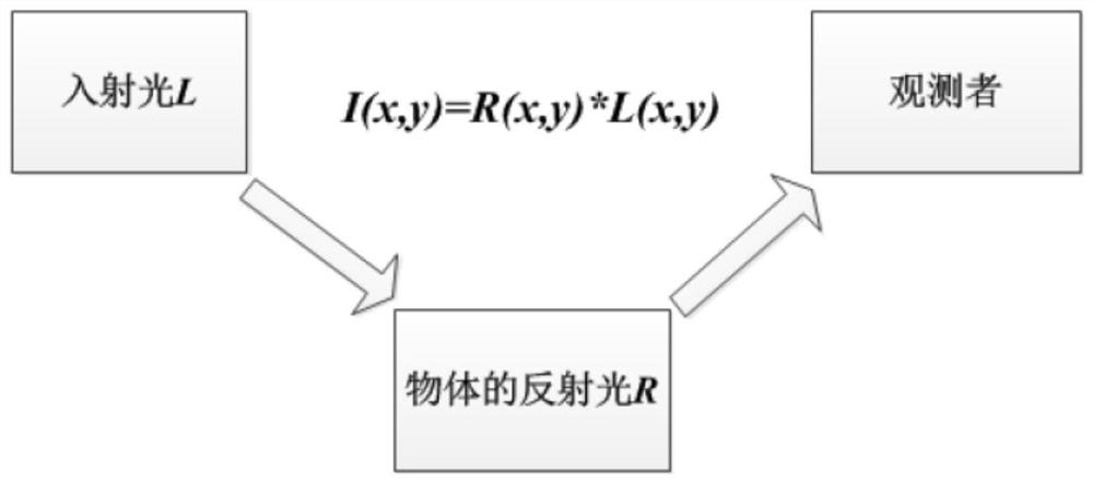
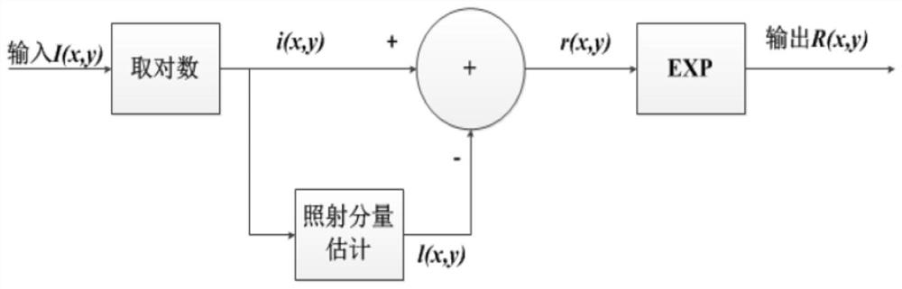
[0034] Step S2, using the Retinex algorithm to enhance the contrast of the anterior segment tomographic image;

[0035] Step S3, performing Gaussian filtering to remove the noise generated after step S2;

[0036] Step S4, through binarization and blob shape analysis to find out the potential corneal area (a is the ideal edge jump, b is the figure obtained by calculating the first derivative of the ideal edge jump);

[0037] Step S5, using the gradient maximum method for the potential corneal area to roughly locate the front and rear surface edges of the cornea;

[0038] Step S6, determining the su...


PUM


Abstract

The invention discloses an anterior segment sectional image feature extraction method based on machine vision, and the method comprises the following steps: collecting an anterior segment sectional image under low-illumination illumination; enhancing the contrast of the anterior segment sectional image by adopting a Retinex algorithm; performing Gaussian filtering to remove noise generated after the step S2; finding a potential corneal region through binarization and blob shape analysis; roughly positioning the edges of the front and rear surfaces of the cornea in the potential cornea region by using a gradient maximum method; determining sub-pixel precision boundaries of the front surface and the rear surface of the cornea through a Gaussian fitting positioning method; according to the solved sub-pixel precision boundaries of the front and rear surfaces of the cornea, finding initial points of the iris and the crystalline lens, and obtaining accurate boundary values of the front and rear surfaces of the cornea, the front surface of the crystalline lens and the front surface of the iris through tracking. The invention has the following advantages and effects that high-precision processing can be performed on the image in a low-illumination imaging mode, and the matching degree and the comfort degree of a patient can be greatly improved.

Description

technical field [0001] The invention relates to the technical field of ophthalmology detection, in particular to a feature extraction method of an anterior segment tomographic image based on machine vision. Background technique [0002] When using a slit lamp or related equipment to collect anterior segment tomographic images, in order to ensure image quality, a high-intensity light source is usually used to irradiate the human eye. Some people who are sensitive to light cannot even cooperate to complete the entire detection, requiring the cooperation of an eye opener, and the eyes cannot be used during the test. Concentrate on the point of gaze, resulting in a decrease in measurement accuracy. Therefore, if the patient's discomfort can be reduced by reducing the intensity of the illumination source, and the image can be processed with high precision even in the low-light imaging mode, the cooperation and comfort of the patient will be greatly improved. Contents of the inv...

Claims


Application Information

Patent Timeline
Patent Type & Authority Applications(China)
IPC IPC(8): G06T7/00G06T7/13G06T7/11
CPCG06T7/0012G06T7/13G06T7/11G06T2207/30041G06T2207/10072
Inventor 王勤美黄锦海邓大辉高蓉蓉梅晨阳曾震海
Owner THE EYE HOSPITAL OF WENZHOU MEDICAL UNIV
Why Eureka
Try Eureka