Medical robot operation video motion amplification method

A technology of medical robot and surgical video, applied in the field of medical robot

Active Publication Date: 2020-11-06
罗雄彪 +3
View PDF4 Cites 0 Cited by
  • Summary
  • Abstract
  • Description
  • Claims
  • Application Information

AI Technical Summary

Problems solved by technology

But this method introduces addition

Method used


Image

  • Medical robot operation video motion amplification method
  • Medical robot operation video motion amplification method
  • Medical robot operation video motion amplification method

Examples


Embodiment Construction

[0048] In order to make the purpose, technical solutions and advantages of the embodiments of the present invention more clear, the technical solutions in the embodiments of the present invention will be clearly and completely described below in conjunction with the accompanying drawings in the embodiments of the present invention. Obviously, the described embodiments It is a part of embodiments of the present invention, but not all embodiments. Based on the embodiments of the present invention, all other embodiments obtained by persons of ordinary skill in the art without making creative efforts belong to the protection scope of the present invention.

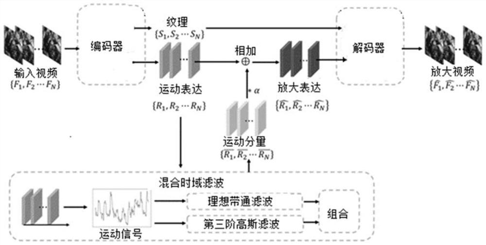
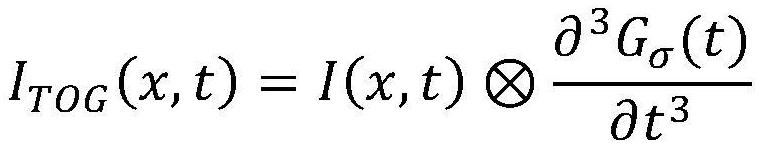
[0049] figure 1 Shown is the flow chart of the video motion amplification method for medical robot surgery proposed by the present invention. The motion amplification method of medical robot surgery video of the present invention is based on Euler motion amplification, and mainly includes three steps: spatial decomposition ba...


PUM


Abstract

The invention provides a medical robot operation video motion amplification method which comprises the following steps: A, performing spatial decomposition on all frames {F1, F2... FN} of an input video to obtain a texture data set {S1, S2... SN} and a shape data set {R1, R2... RN}, and N is a positive integer greater than 1; B, performing hybrid time domain filtering processing on the data set {S1, S2... SN} of the texture and the data set {R1, R2... RN} of the shape; and C, amplifying the data set obtained in the step B and recombining the video. According to the medical robot operation video motion amplification method, based on a brand-new operation vision enhancement algorithm of video motion amplification, pulsation of nerve blood vessel bundles in an operation video is amplified. Through the video motion amplification algorithm, a doctor can easily observe and obtain the position of the nerve blood vessel bundle through naked eyes, so that the nerve blood vessel bundle is prevented from being accidentally injured in the operation process.

Description

technical field [0001] The invention belongs to the field of medical robots, and in particular relates to a method for amplifying the motion of a medical robot operation video. Background technique [0002] The existing minimally invasive surgery has replaced the traditional open surgery and has become one of the mainstream surgical methods because of its fast recovery and few complications. But minimally invasive surgery also has some drawbacks. In traditional open surgery, surgeons are able to use visual and tactile perception to localize neurovascular bundles in the surgical environment. However, in minimally invasive surgery, doctors cannot use the sense of touch to sense the neurovascular bundles in the surgical environment, and the naked eye cannot locate the neurovascular bundles in the endoscopic video, so it is easy to accidentally injure the neurovascular bundles during minimally invasive surgery bundle. Although some traditional interventional imaging methods c...

Claims


Application Information

Patent Timeline
IPC IPC(8): G06T3/40H04N5/14
CPCG06T3/4046H04N5/148
Inventor 罗雄彪万英
Owner 罗雄彪
Why Eureka
Try Eureka