Method for determining and verifying screw channel for bone surgery based on three-dimensional modeling

A three-dimensional modeling and verification method technology, applied in computer-aided surgery, computer-aided planning/modeling, medical science, etc., can solve the problems of difficult screw paths, poor intraoperative guidance, and high error rates, and achieve application Low cost, improved nail placement accuracy, and easy popularization

Pending Publication Date: 2020-12-04
ZIGONG NO 4 PEOPLES HOSPITAL
View PDF10 Cites 1 Cited by
  • Summary
  • Abstract
  • Description
  • Claims
  • Application Information

AI Technical Summary

Problems solved by technology

At present, it is difficult to quickly obtain the screw passages of the sacral and atlantoaxial vertebrae clinically. The position of the screw is often determined by plane CT or X-ray film. The error rate is high and the intraoperative guidance is poor. The determination of the screw insertion point is often done by the clinician. The experience is strong, subjectivity is strong, and the technology is not easy to popularize at the grassroots level

Method used


Image

  • Method for determining and verifying screw channel for bone surgery based on three-dimensional modeling
  • Method for determining and verifying screw channel for bone surgery based on three-dimensional modeling
  • Method for determining and verifying screw channel for bone surgery based on three-dimensional modeling

Examples


Embodiment 1

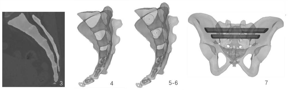
[0034] like figure 1 As shown, a method for determining and verifying the sacral transverse screw channel based on three-dimensional modeling includes the following steps:

[0035] S1. Obtain 256 rows of dual-source CT scan data of the sacrum. The scan parameters are: voltage 120kV, current 150mA, slice thickness 0.625mm, scan without interval, scan matrix 512X512 pixels, and save it in Dicom format;

[0036] S2, importing the original Dicom data into the three-dimensional modeling software Mimics 22.0 (Materialise company, Belgium);

[0037] S3. Create a new bone mask in "new mask". In the middle layer of the coronal plane, use "split mask" to separate the sacrum mask from the upper and lower adjacent vertebral body masks, and "region growing" to separate the two-dimensional mask of the sacrum independently Come out and fill the bone completely in the mask;

[0038] S4. Reconstruct the three-dimensional model of the sacrum based on the above-mentioned two-dimensional mask "...

Embodiment 2

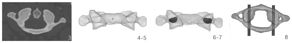
[0043] like figure 2 As shown, a method for determining and verifying the pedicle screw channel of the atlas without inclination angle based on three-dimensional modeling includes the following steps:

[0044] S1. Obtain 256 rows of dual-source CT scan data of the sacrum. The scan parameters are: voltage 120kV, current 150mA, slice thickness 0.625mm, scan without interval, scan matrix 512X512 pixels, and save it in Dicom format;

[0045] S2, importing the original Dicom data into the three-dimensional modeling software Mimics 22.0 (Materialise company, Belgium);

[0046] S3. Create a new bone mask in "newmask", and use "split mask" to separate the atlas mask from the upper and lower adjacent vertebral body masks in the middle layer of the coronal plane, and use "region growing" to separate the two-dimensional mask of the atlas Segment it and fill the bone completely in the mask;

[0047] S4. Reconstruct the three-dimensional model of the atlas based on the above-mentioned t...

Embodiment 3

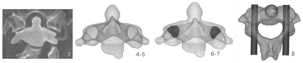
[0053] like image 3 As shown, a method for determining and verifying the pedicle screw channel of the axis without inclination angle based on three-dimensional modeling includes the following steps:

[0054] S1. Obtain 256 rows of dual-source CT scanning data of the axis vertebrae. The scanning parameters are: voltage 120kV, current 150mA, slice thickness 0.625mm, no interval scanning, scanning matrix 512X512 pixels, and save in Dicom format;

[0055] S2, importing the original Dicom data into the three-dimensional modeling software Mimics 22.0 (Materialise company, Belgium);

[0056]S3. Create a new bone mask in "new mask". In the middle layer of the coronal plane, use "split mask" to separate the mask of the axis from the upper and lower adjacent vertebrae. "region growing" separate the two-dimensional mask of the axis Segment it independently, and fill the bone completely in the mask;

[0057] S4. Reconstruct the three-dimensional model of the axis based on the above-men...


PUM


Abstract

The invention discloses a method for determining and verifying a screw channel for bone surgery based on three-dimensional modeling. The method comprises the steps of firstly, obtaining original CT scanning data of a bone, storing the original CT scanning data in a Dicom format, importing the original Dicom data into three-dimensional modeling software, building a two-dimensional mask of a bone needing to be operated, separating the mask of the bone needing to be operated from a mask of an adjacent bone, independently separating out the two-dimensional mask of the bone needing to be operated,and filling the mask with bone substances completely; reconstructing a three-dimensional model of the vertebral body based on the two-dimensional mask of the bone; in a high perspective mode of the three-dimensional model of the vertebral body, obtaining the form of the screw channel on the vertebral body; and projecting a vertebral arch channel to a median coronal plane or sagittal plane to obtain a screw channel projection, fitting the maximum inscribed circle of the screw channel, recording the radius of the inscribed circle, and obtaining the position of the circle center projected to thesurface of the vertebral body, namely the optimal screw feeding point. By establishing the three-dimensional model, the screw channel is rapidly extracted, the screw placement accuracy is high, the cost is low, and popularization is facilitated.

Description

technical field [0001] The invention relates to the field of preoperative planning for spinal surgery, in particular to a method for determining and verifying a channel for orthopedic surgery based on three-dimensional modeling. Background technique [0002] In spinal surgery, screw implantation is one of the key techniques of surgery. At present, it is difficult to quickly obtain the screw passages of the sacral and atlantoaxial vertebrae clinically. The position of the screw is often determined by plane CT or X-ray film. The error rate is high and the intraoperative guidance is poor. The determination of the screw insertion point is often done by the clinician. It is highly subjective, and the technology is not easy to popularize at the grassroots level. Contents of the invention [0003] In view of the above problems, the present invention provides a method for determining and verifying screw placement channels in orthopedic surgery based on three-dimensional modeling....

Claims


Application Information

Patent Timeline
Patent Type & Authority Applications(China)
IPC IPC(8): A61B34/10
CPCA61B34/10A61B2034/108A61B2034/104
Inventor 吴超邓佳燕潘建胡海刚王翔宇
Owner ZIGONG NO 4 PEOPLES HOSPITAL
Why Eureka
Try Eureka