Subretinal fluid drainage device and collector

A retina and collector technology, applied in the field of medical devices, can solve problems such as poor safety, difficulty in evaluating drainage volume, and increased infection, and achieve the effects of ensuring safety, preventing sliding and sliding off the sclera, and facilitating laboratory testing

Pending Publication Date: 2020-12-08
BEIJING TONGREN HOSPITAL AFFILIATED TO CAPITAL MEDICAL UNIV
View PDF3 Cites 0 Cited by
  • Summary
  • Abstract
  • Description
  • Claims
  • Application Information

AI Technical Summary

Problems solved by technology

[0003] Most of the functions of the subretinal fluid drainage devices currently on the market are to realize retinal drainage. For example, Chinese patent (CN203370018U) discloses a subretinal fluid drainage valve composed of a drainage tube, a negative pressure valve, and a fixing ring. Previously, a 5ml needle was needed to puncture the scleral wall, the drainage valve and the eyeball wall were sutured and fixed during the operation, and the negative pressure valve was used to continuously drain the subretinal fluid, which was taken out after 2-3 weeks. It had the following defects: 5ml needle puncture increased retinal risk of sub- or retinal hemorrhage; need for secondary surgery; increased risk of infection; possibility of drain valve falling into the eye or sub- / outside the conjunctiva; rejection
Another example is that Chinese patent (CN201668586U) discloses a retinal liquid discharge device consisting of a positioning tube, a liquid discharge needle, and a push rod. It has the following drawbacks: the small needle does not ensure a steady outflow of subretinal fluid
Another example is that the Chinese patent (CN209122625U) discloses a puncture needle comprising a puncture needle, a needle seat, and a three-way structure. Drainage is carried out by kneading a flexible cystic structure. It has the following defects: it is difficult to evaluate the drainage volume, and the operation process In the middle, the condition of the fundus cannot be grasped in time
Therefore, the subretinal fluid drainage device in the prior art is less safe, and there are certain hidden dangers that make the drainage difficult to complete smoothly, and lack the function of collecting subretinal fluid, making it difficult to provide useful information for disease diagnosis and identification

Method used


Image

  • Subretinal fluid drainage device and collector
  • Subretinal fluid drainage device and collector
  • Subretinal fluid drainage device and collector

Examples


Embodiment 1

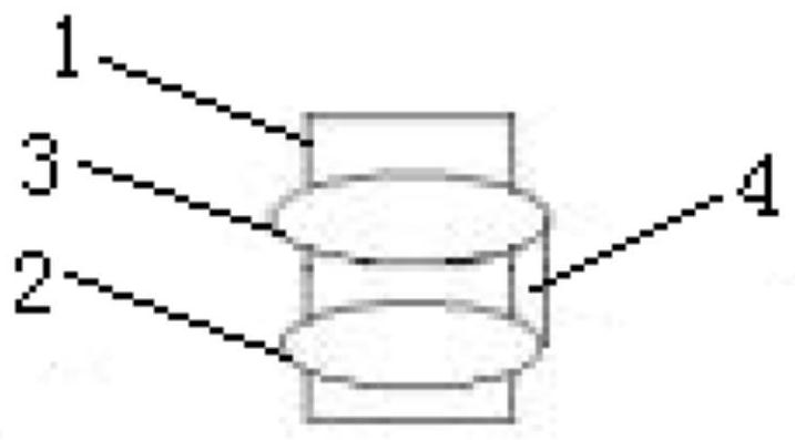
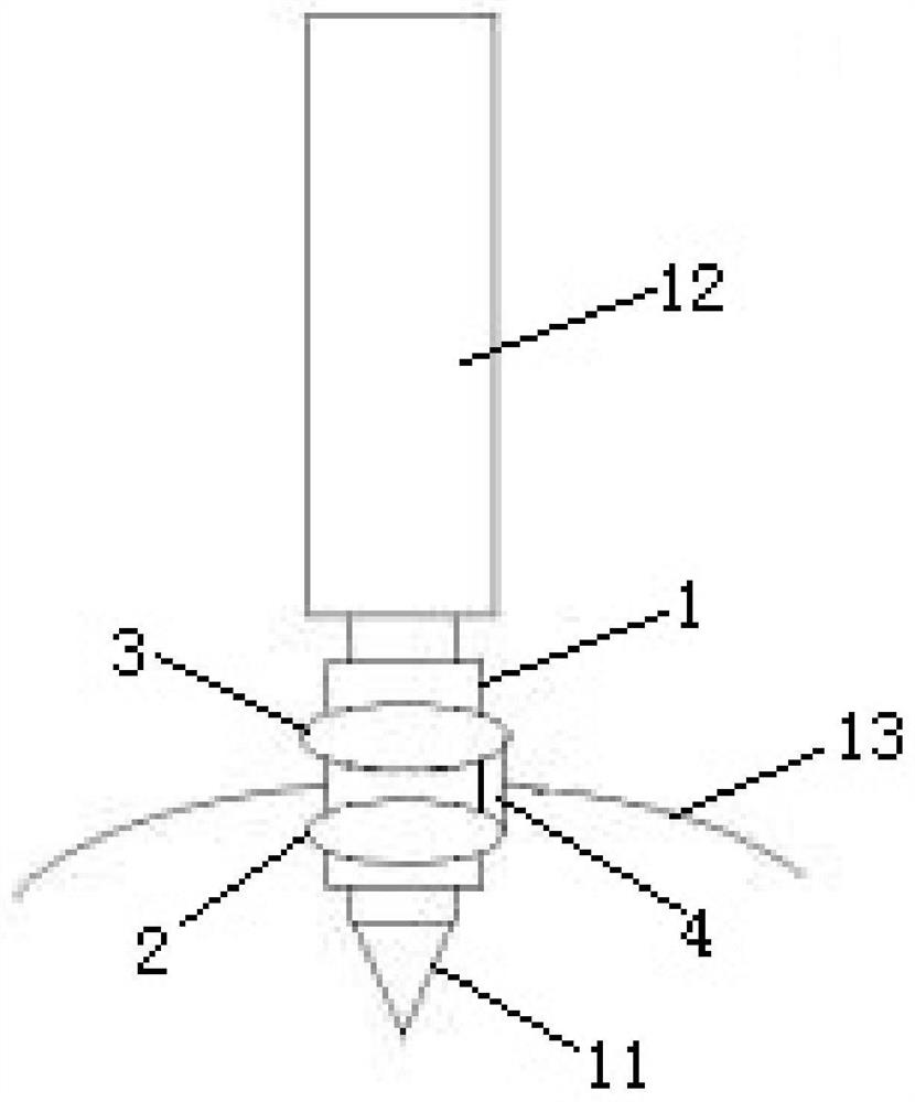
[0028] like figure 1 As shown, the present embodiment provides a retinal under fluid drainage device including a sleeve 1, and the sleeve 1 is provided with a first capsule 2 and a second capsule 3, a first capsule 2 and a second capsule. The body 3 is provided on the surface of the sleeve 1, and the spacing between the first capsule 2 and the second capsule 3 is small. The first capsule 2 and the second capsule 3 may be charged into the liquid or gas, respectively, and the first capsule 2 and the second capsule 3 are in a bulk state; the first capsule 2 and the second capsule 3 After the liquid or gas is exhausted, the first capsule 2 and the second capsule 3 are in a contracted state.

[0029] When the retinal is discharged, the first capsule 2 and the second capsule 3 are respectively located on both sides of the sclera 13, and the first capsule 2 and the second capsule 3 are in a state in which the casing 1 is prevented. Slippery. When used in specifically, the first capsule 2...

Embodiment 2

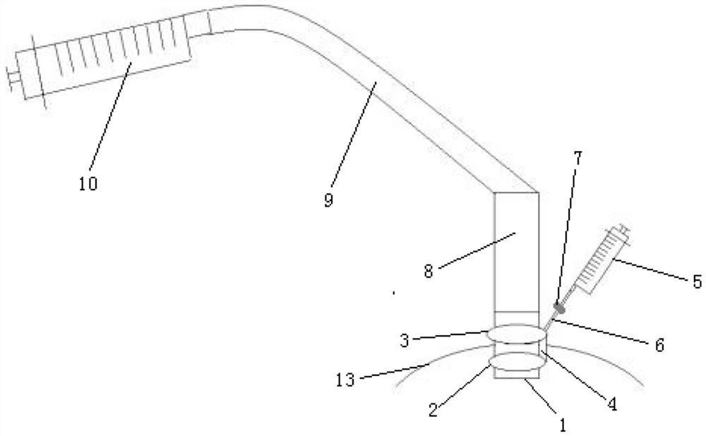
[0035] likefigure 2 and image 3 , The present embodiment provides a collection of subretinal fluid, comprising drainage means, needle, and collecting pipe connection apparatus, said drainage device comprising a casing 1, the sleeve 1 is provided with a first balloon 2 and 3 a second balloon, the first balloon and the second balloon 2 is provided on a surface of the sleeve 3, and a smaller distance between the first balloon and the second balloon 32. The puncture needle and the cannula 1 match with each other, the cannula 1 is connected with the collection tube by connecting means. Needle cannula 1 can be installed in the puncture needle after breaking the sclera 13, the puncture needle is removed from the casing 1, and then install the connecting pipe, subretinal fluid collection. The length of the puncture needle is 3.5mm, the length of the sleeve 1 is 2.5-3mm.

[0036] The first and the second capsule 2 capsule 3 may be charged with liquid or gas, respectively, the first balloon...


PUM


Abstract

The invention discloses a subretinal fluid drainage device and a collector. The subretinal fluid drainage device comprises a sleeve, and the sleeve is provided with a first bag body and a second bag body. During subretinal fluid drainage, the first bag body and the second bag body are located at two sides of the sclera respectively, and the first bag body and the second bag body are both in bulgedstates, so that positioning of the sleeve is realized, the sleeve is prevented from sliding and sliding away from the sclera, the safety of subretinal fluid drainage is ensured, and the subretinal fluid drainage can be smoothly carried out.

Description

Technical field [0001] The present invention belongs to the technical field of medical device, and more particularly to a retinal under fluid drainage device and a collector. Background technique [0002] In the clinical work of ophthalmology, retinal disengagement (bleeding or pore sources) is a serious symptom of vision, these symptoms can be found in some eye trauma, inflammation, and internal eye surgery, and remove the retina as a test specimen, available In further laboratory testing, it is a potential disease diagnosis, identifying diagnosis and development direction. [0003] At present, the function of the retinal liquid drainage device on the market is mostly to achieve retinal discharge, such as the Chinese patent (CN203370018U) discloses a retinal subtracheal drainage valve composed of a drain tube, a negative pressure valve, a fixed ring, and an implantation. It is necessary to use 5ml needle to pierce the sclera wall, which is fixed by the drainage valve and the eye...

Claims


Application Information

Patent Timeline
Patent Type & Authority Applications(China)
IPC IPC(8): A61F9/007A61B10/00
CPCA61B10/0045A61B2010/0067A61F9/007A61F2009/00863
Inventor 彭晓燕呼风曹鑫杨
Owner BEIJING TONGREN HOSPITAL AFFILIATED TO CAPITAL MEDICAL UNIV
Why Eureka
Try Eureka