Method for quantitatively evaluating proliferation of tumor cells transplanted into zebra fish embryos

A technology for tumor cell proliferation and zebrafish embryos, which is applied in the field of biotechnology, and can solve the problems that still exist, cannot realize direct observation of tumor cell proliferation, and cause large damage to animal embryo cells.

Pending Publication Date: 2020-12-18
NANJING YSY BIOTECH CO LTD
View PDF7 Cites 3 Cited by
  • Summary
  • Abstract
  • Description
  • Claims
  • Application Information

AI Technical Summary

Problems solved by technology

However, this method also has obvious disadvantages, that is, the fluorescent dyes will not increase with the proliferation of cells, and the fluorescent dyes on non-proliferated cell fragments still exist.
Therefore, direct observation of tumor cell proliferation cannot be achieved using this labeling method.
[0008] In addition, the allogeneic cell transplantation causes great damage to animal embryo cells. In practice, a considerable number of embryos die during the subsequent development of the embryo due to mechanical damage during cell transplantation, so they cannot be used as evaluation objects.

Method used


Image

  • Method for quantitatively evaluating proliferation of tumor cells transplanted into zebra fish embryos
  • Method for quantitatively evaluating proliferation of tumor cells transplanted into zebra fish embryos

Examples


Embodiment 1

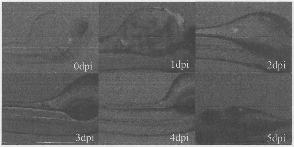
[0024] Example 1: Quantitative evaluation of colonization and proliferation of human colon cancer cell HCT116 and human non-small cell lung cancer cell A549 transplanted into zebrafish embryos

[0025] Materials and methods (experimental methods in the embodiments, if no special instructions, are conventional methods)

[0026] Cell line: human colon cancer cell HCT116, human non-small cell lung cancer cell A549

[0027] Complete medium formula for HCT116 cells: one bag of DMEM powder, high glucose (Gibco), another weighed 3.7g NaHCO 3 (Sigma), add ultrapure water (18.2 megohm) to make up to 1L, after filtration, perform autoclave sterilization at 121°C for 30min, and filter in an ultra-clean bench. Add 50 mL of fetal bovine serum (PAN) and 5 mL of penicillin-streptomycin (10,000 U / mL) to 500 mL of the filtered liquid, mix well, divide into 50 mL centrifuge tubes, seal with parafilm, and store in a refrigerator at 4 °C spare.

[0028] Medium for A549 cells: Ham's F-12K (Kai...


PUM

PropertyMeasurementUnit
radiusaaaaaaaaaa

Abstract

The invention belongs to the technical field of biology, and relates to a method for quantitatively evaluating proliferation of tumor cells transplanted into zebra fish embryos. The method is characterized by comprising the following steps of: performing fluorescence labeling on the tumor cells before injection; selecting the zebra fish embryos carrying the fluorescence tumor cells for further culture one day after injection; and amplifying human tumor cell genes by a quantitative PCR method 3 to 7 days after injection to evaluate the colonization and proliferation conditions of the human tumor cells in the zebra fish embryos. The injection period of the tumor cells subjected to fluorescence labeling in advance is the period of 48 hours after fertilization, and the injection part is a perivitelline space area. The injection volume of each embryo is 10 nL, and the number of the injected tumor cells is 300 to 500. During quantitative evaluation, a plurality of embryos are selected as a group generally, and the content of the human genes is detected by quantitative PCR. The period of 3 to 7 days after injection is an effective time window for evaluating whether the human tumor cells are proliferated in the embryos. The method is beneficial to the practicability of a PDX (patient-derived cell transplantation) model of zebra fish.

Description

technical field [0001] The invention belongs to the field of biotechnology, and relates to a method for quantitatively evaluating the proliferation of tumor cells transplanted into zebrafish embryos, which is characterized in that the tumor cells are fluorescently labeled before injection, and the zebrafish embryos carrying fluorescent tumor cells are selected one day after injection Further culture, and the human tumor cell colonization and proliferation in zebrafish embryos were evaluated by quantitative PCR method during the window period of 3 to 7 days after injection (equivalent to 5 to 9 days after fertilization). [0002] technical background [0003] It is generally believed that the occurrence of tumors is due to the cumulative effect of multiple gene mutations. Due to the diversity of gene mutations in cancer patients, existing tumor-targeted drugs can only be curative for cancer patients carrying a specific type of gene mutation. Therefore, for different tumor pat...

Claims


Application Information

Patent Timeline
Patent Type & Authority Applications(China)
IPC IPC(8): C12Q1/6851
CPCC12Q1/6851C12Q2531/113C12Q2563/107
Inventor 不公告发明人
Owner NANJING YSY BIOTECH CO LTD
Why Eureka
Try Eureka