Method for evaluating collateral vessels and tissue function based on cerebral blood flow

A technique of cerebral blood flow and cerebral blood flow, applied in the direction of blood flow measurement, catheterization, cardiac catheterization, etc., can solve the problems of difficulty in visualization of tertiary collateral circulation and evaluation of ischemic penumbra, and achieve the goal of evaluating prognosis Effect

Active Publication Date: 2021-01-26
THE FIRST MEDICAL CENT CHINESE PLA GENERAL HOSPITAL +1
View PDF5 Cites 2 Cited by
  • Summary
  • Abstract
  • Description
  • Claims
  • Application Information

AI Technical Summary

Problems solved by technology

Cerebral collateral circulation is divided into three levels, among which the third-level collateral circulation is related to the ischemic penumbra, which often refers to the rapid establishment of small arteries after the occurrence of ischemic stroke to supply blood to the ischemic penumbra. This kind of tertiary collateral circulation imaging can well refle

Method used


Image

  • Method for evaluating collateral vessels and tissue function based on cerebral blood flow
  • Method for evaluating collateral vessels and tissue function based on cerebral blood flow
  • Method for evaluating collateral vessels and tissue function based on cerebral blood flow

Examples


Embodiment Construction

[0025] In order to facilitate those of ordinary skill in the art to understand and implement the present invention, the present invention will be described in further detail below in conjunction with the examples. It should be understood that the implementation examples described here are only used to illustrate and explain the present invention, and are not intended to limit the present invention.

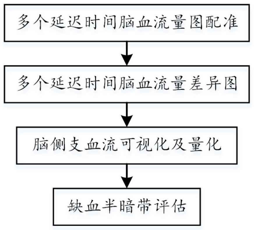
[0026] A method for assessing collateral vessel and tissue function based on cerebral blood flow, comprising the following steps:

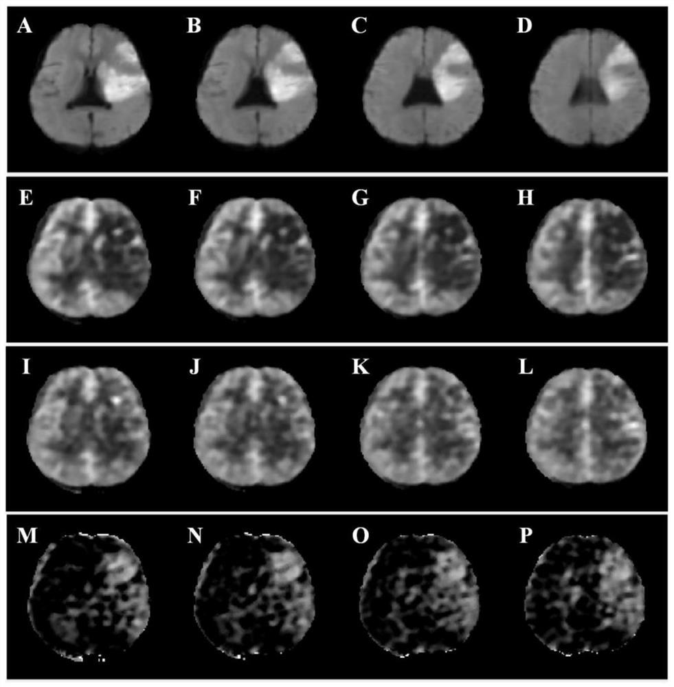
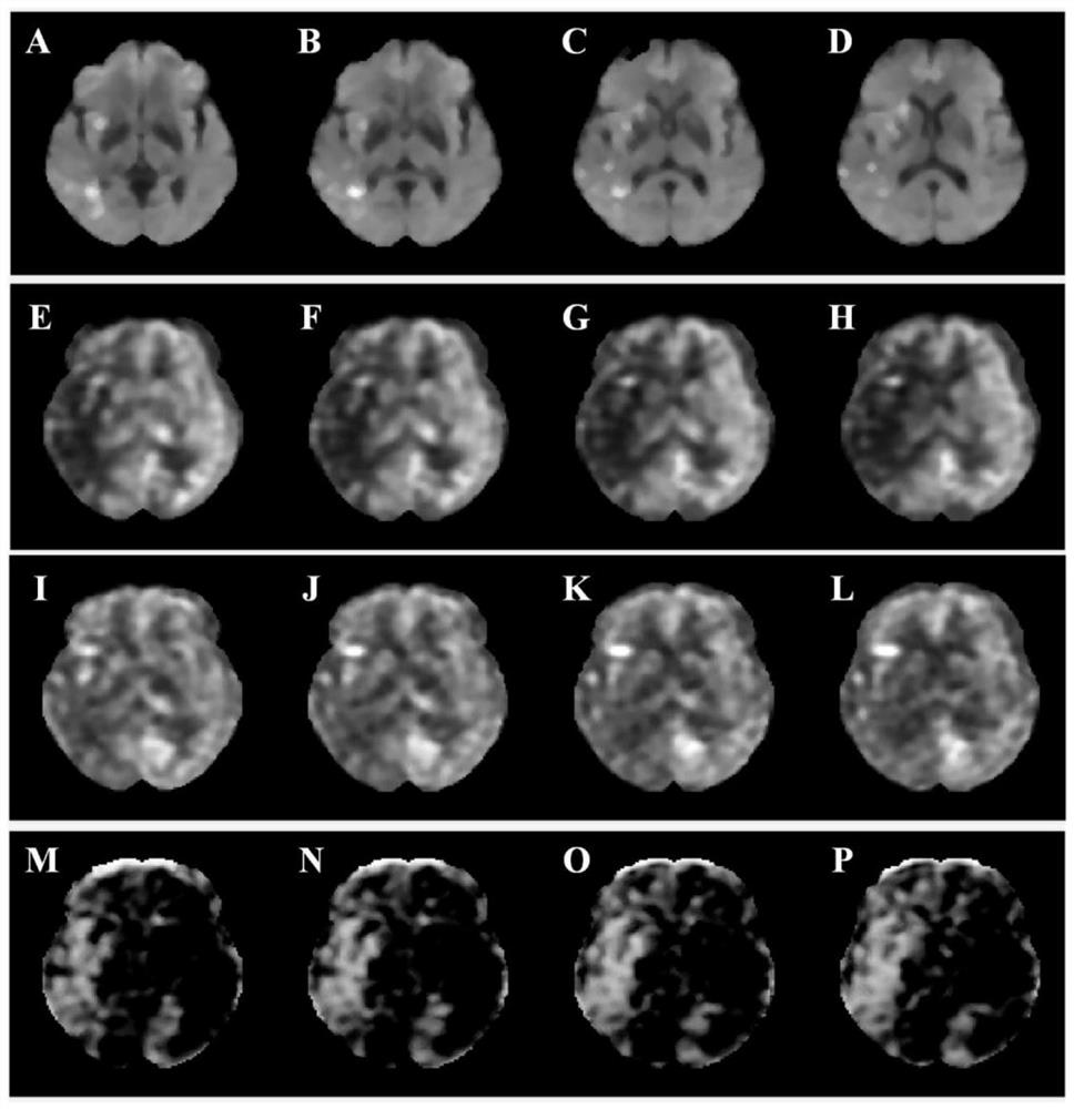
[0027] Step 1, registration of cerebral blood flow maps of multiple delay times in patients diagnosed with large artery occlusive infarction, so that the cerebral blood flow maps of each delay time are mapped one by one on the spatial anatomical structure.

[0028] In the clinical magnetic resonance examination of large artery occlusive infarction, the scanning order of the imaging sequence is generally structural imaging first (T1 / T2-FLAIR), followed ...


PUM


Abstract

The invention discloses a method for evaluating collateral vessels and tissue function based on cerebral blood flow, which comprises the following steps of: firstly, registering cerebral blood flow graphs with multiple delay times of a patient diagnosed with major artery occlusion infarction to realize one-to-one correspondence on a spatial anatomical structure; secondly, removing cerebral shellsand extracerebral structures of the cerebral blood flow graphs with multiple delay times after registration, and calculating cerebral blood flow difference graphs with multiple delay times; then, visualizing cerebral collateral blood circulation conditions, and quantifying collateral blood flow related information, such as collateral blood flow distribution range, velocity, flow and the like; andfinally, by combining with the infarction core area of the patient diagnosed with the major artery occlusion infarction, evaluating the ischemic penumbra, such as the size, volume and position of theischemic penumbra.

Description

technical field [0001] The invention belongs to the technical field of magnetic resonance imaging and medical image processing, and in particular relates to a collateral vessel and tissue function evaluation method based on cerebral blood flow. Background technique [0002] The currently widely used method is to use the perfusion imaging method of contrast agent injection to obtain the pixel-level contrast agent inflow-time curve, so as to obtain multiple parameters that can reflect hemodynamics, such as average transit time, time to peak, Peak reaction time, etc., determine the severe hypoperfusion area by limiting the time threshold, so as to form a mismatch with the brain tissue death area to reflect the ischemic penumbra. However, the limitations of this method are obvious. Firstly, it may not be the ischemic penumbra in the true sense to reflect severe hypoperfusion simply by setting the time threshold. It also includes benign hypoperfusion areas. Secondly, the above-me...

Claims


Application Information

Patent Timeline
IPC IPC(8): A61B5/02A61B5/026
CPCA61B5/0263A61B5/02007A61B5/72
Inventor 娄昕周欣吕晋浩邓鹤肖洒孙献平叶朝辉
Owner THE FIRST MEDICAL CENT CHINESE PLA GENERAL HOSPITAL
Why Eureka
Try Eureka