PET image reconstruction method and system

An image reconstruction and imaging system technology, applied in the field of medical imaging, can solve the problems of insufficient sample data, affecting PET image quality and quantitative analysis, etc., to enhance local smoothness, preserve tumor edge contrast, and improve PET image quality. and tumor detection effect

Pending Publication Date: 2021-02-12
JIANGSU SINOGRAM MEDICAL TECH CO LTD
View PDF0 Cites 0 Cited by
  • Summary
  • Abstract
  • Description
  • Claims
  • Application Information

AI Technical Summary

Problems solved by technology

However, PET acquisition is usually limited by the insufficient amount of collected sample data, so a common problem of ML methods is data ill-conditioning, that is, the reconstructed image is very sensiti

Method used


Image

  • PET image reconstruction method and system
  • PET image reconstruction method and system
  • PET image reconstruction method and system

Examples


Embodiment 1

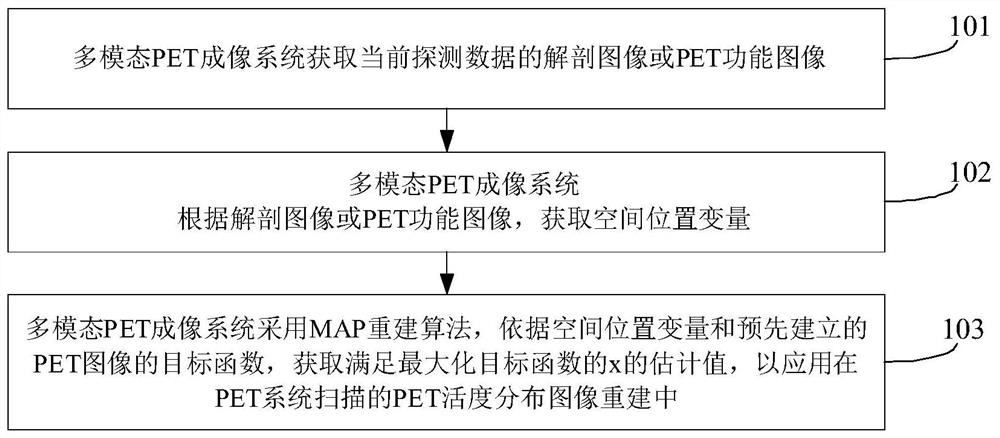
[0061] Such as figure 1 as shown, figure 1 It shows a flowchart of a method for PET image reconstruction provided by an embodiment of the present invention. The execution subject of the method of this embodiment may be a multi-modal PET imaging system connected by communication with PET equipment. The method of this embodiment may include the following Methods:

[0062] Step 101, the multimodal PET imaging system acquires anatomical images or PET functional images of current detection data;

[0063] In this embodiment, the multimodal PET imaging system may be an imaging system integrated with other modality imaging functions, and then may generate anatomical images or PET functional images according to the acquired current detection data.

[0064] For example, the anatomical image in this embodiment can be an MRI image or a CT image; in a specific application, the MRI or CT imaging device in the multimodal PET imaging system acquires the detection data of the current examine...

Embodiment 2

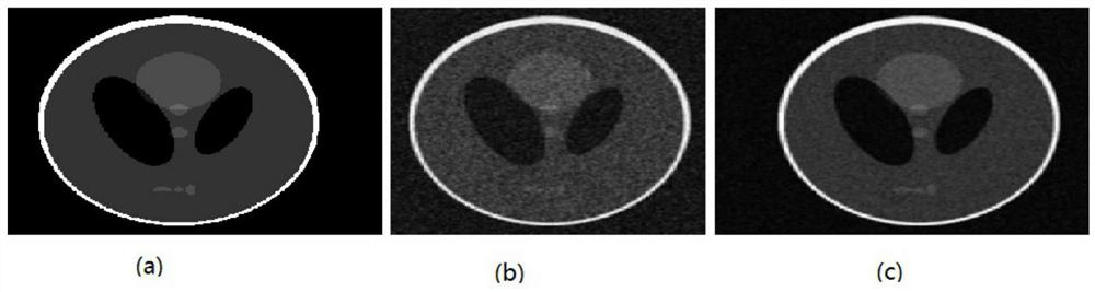
[0080] The present invention proposes a reconstruction algorithm using anatomical prior or functional prior in a multi-modal PET imaging system. While enhancing the local smoothness of the PET image, the contrast of the tumor edge is preserved as much as possible, and the PET image is simultaneously improved. quality and tumor detectability purposes. The specific steps of this method are as follows:

[0081] Step S1, the PET acquisition process can be modeled as the following formula:

[0082]

[0083] In the formula, y=[y 1 ,y 2 ,...,y N ] T Indicates the detected data, and N indicates the dimension of the detected data. For listmode reconstruction, N is the number of detected cases; for sinogram-based reconstruction, N is the size of the sinogram; if the acquisition case includes time resolution TOF (Time of Flight) information, N should also include the dimension of time resolution discretization.

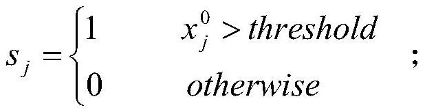
[0084] x=[x 1 ,x 2 ,...,x M ] T Represents the unknown PET rad...


PUM


Abstract

The invention relates to a PET image reconstruction method and system. The method comprises the steps that 101, a multi-mode PET imaging system obtains a PET function image or an anatomical image of acurrent imaging object; 102, the multi-mode PET imaging system obtains a spatial position variable gamma jk according to the anatomical image or the PET functional image; and 103, the multi-mode PETimaging system adopts an MAP reconstruction algorithm, and obtains an estimated value of x satisfying a maximized objective function according to the spatial position variable gamma jk and the pre-established objective function of the PET image. The invention provides a method for extracting smooth priori by using a functional image or an anatomical image, functional boundary information is adaptively extracted as much as possible, the tumor edge contrast is reserved while the image noise is reduced, and finally, the purpose of improving the PET image quality and the tumor detection performance is achieved.

Description

technical field [0001] The invention relates to the technical field of medical imaging, in particular to an image reconstruction method and system which can be used in a positron emission computed tomography imaging system. Background technique [0002] Positron Emission Tomography PET (Positron Emission Tomography) is a high-end nuclear medicine imaging diagnostic equipment. In practice the use of radionuclides (eg 18 F. 11 C, etc.) Label the metabolites and inject the nuclides into the human body, and then perform functional metabolic imaging on the patient through the PET system to reflect the metabolic activities of life, so as to achieve the purpose of diagnosis. Currently commercial positron emission tomography PET is usually integrated with other modal imaging systems, such as computed tomography CT (Computed Tomography) or magnetic resonance imaging MRI (Magnetic Resonance Imaging), to achieve the purpose of simultaneously imaging the patient's anatomical structure...

Claims


Application Information

Patent Timeline
IPC IPC(8): G06T17/05G06T5/00G06T7/11G16H30/20
CPCG06T5/002G06T17/05G06T2207/10081G06T2207/10088G06T2207/10104G06T2207/20076G06T2207/20192G06T2207/30096G06T7/11G16H30/20
Inventor 李楠
Owner JIANGSU SINOGRAM MEDICAL TECH CO LTD
Why Eureka
Try Eureka