Medical image classification method and device based on multi-view learning and depth supervision auto-encoder

A medical imaging and autoencoder technology, applied in image analysis, image enhancement, image data processing, etc., can solve problems such as parameter adjustment and function selection sensitivity

Inactive Publication Date: 2021-03-12
CENT SOUTH UNIV
View PDF5 Cites 4 Cited by
  • Summary
  • Abstract
  • Description
  • Claims
  • Application Information

AI Technical Summary

Problems solved by technology

However, SVM is sensitive to parameter tuning and the choice of function

Method used


Image

  • Medical image classification method and device based on multi-view learning and depth supervision auto-encoder
  • Medical image classification method and device based on multi-view learning and depth supervision auto-encoder
  • Medical image classification method and device based on multi-view learning and depth supervision auto-encoder

Examples


Embodiment 1

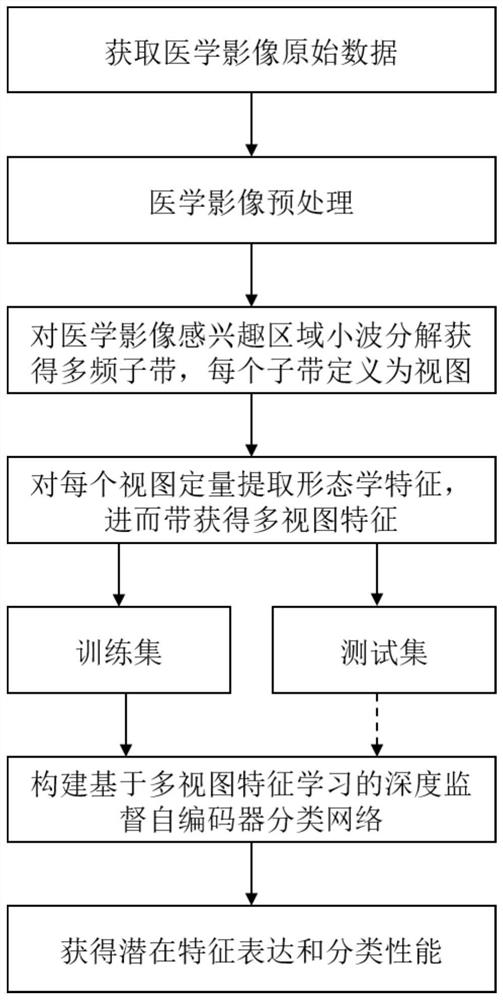
[0075] see figure 1 The image classification method based on multi-morphological multi-task feature selection provided by this embodiment contains the following steps:

[0076] Step 1: Obtain the CT image of the subject (in this embodiment, CT is the image), and first perform preprocessing on the image of each subject, specifically: use the dcm2nii software package to convert the medical image of each case Reconstructed into a 3D image; then the 3D image was preprocessed using the 3D U-Net model to extract the lung parenchyma of the 3D image; in order to overcome the difference between sample thickness variations, the volumetric data of the lung parenchyma were resampled by B-spline interpolation Voxel resolution of 1mm x 1mm x 1mm.

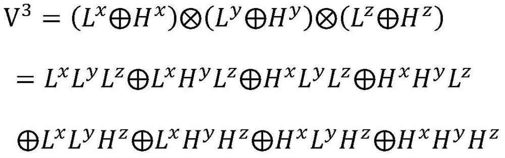
[0077] Step 2: Perform wavelet decomposition on the region of interest of the medical image preprocessed in step 1 to obtain multi-frequency subbands;

[0078] Each segmented volume is textured using 3D-WT to capture eight distinct frequency su...

Embodiment 2

[0126] This embodiment discloses a medical image classification device based on multi-view learning and deep supervision autoencoder, including the following modules:

[0127] The image wavelet transform module is used to perform step 1: use wavelet transform to perform wavelet decomposition on the region of interest of each image, and then obtain multiple subbands in different frequency domains, and each subband is defined as a view.

[0128] The multi-view feature extraction module is used to perform step 2: quantitatively extract 93 morphological features for each view, and then obtain multi-view features;

[0129] The classifier construction and training module is used to perform step 3: construct a deep supervised self-encoder classification network based on multi-view feature learning, input multi-view morphological features to the encoder module, and then obtain high-order potential expressions of multi-view features , and then input the potential expression into the en...

Embodiment 3

[0133] This embodiment discloses an electronic device, including a memory and a processor, where a computer program is stored in the memory, and when the computer program is executed by the processor, the processor implements the method described in Embodiment 1 .


PUM


Abstract

The invention discloses a medical image classification method and device based on multi-view learning and a deep supervision auto-encoder, and the method comprises the following steps: 1, carrying outthe wavelet decomposition of a region of interest of a medical image, and obtaining a multi-frequency sub-band; step 2, defining each sub-band as a view, and quantitatively extracting an image omicsfeature from each view so as to obtain a multi-view feature; step 3, constructing a classification network of a deep supervision auto-encoder based on multi-view feature learning, and training the classification network based on morphological multi-view feature vectors of the image samples and classification tags thereof to obtain a trained classification model; and step 4, classifying the imageswith unknown classification labels based on the trained classification model. According to the invention, the classification accuracy of medical images can be improved.

Description

technical field [0001] The present invention specifically relates to a medical image classification method and device based on multi-view learning and deep supervised self-encoder. Background technique [0002] Medical imaging mainly includes X-ray, computed tomography (CT), positron emission tomography (PET), ultrasound, magnetic resonance imaging (MRI), etc. With the continuous development and progress of medical imaging technology and computer technology, in recent years, medical image classification has become a very important tool in clinical disease diagnosis and medical research. [0003] In many practical problems, the same thing can be described from many different approaches or different angles, and such multiple descriptions constitute multiple views of the same thing. Multi-view can represent different feature sets of data; it can represent the source of data; it can also be used to represent different relationships between data. Multi-view data is ubiquitous i...

Claims


Application Information

Patent Timeline
Patent Type & Authority Applications(China)
IPC IPC(8): G06K9/32G06K9/62G06N3/04G06T5/00G06T5/40G06T7/11G06T7/40
CPCG06T5/002G06T7/11G06T7/40G06T5/40G06T2207/10081G06V10/25G06N3/045G06F18/241G06F18/214
Inventor 王建新成建宏刘军赵伟刘锦
Owner CENT SOUTH UNIV
Why Eureka
Try Eureka