Retinal vessel image segmentation method based on improved UNet + +

A retinal blood vessel and image segmentation technology, applied in the field of medical image processing, can solve the problem of loss of details in segmentation results, and achieve the effect of solving the loss of details, enhancing the generalization ability, and improving the efficiency of use

Active Publication Date: 2021-03-16
KUNMING UNIV OF SCI & TECH
View PDF8 Cites 12 Cited by
  • Summary
  • Abstract
  • Description
  • Claims
  • Application Information

AI Technical Summary

Problems solved by technology

[0004] The invention provides a retinal blood vessel image segmentation method based on improved UNet++, fully uses features of different scales to solve the problem of loss of details in the segmentation results, and achieves better segmentation performance

Method used


Image

  • Retinal vessel image segmentation method based on improved UNet + +
  • Retinal vessel image segmentation method based on improved UNet + +
  • Retinal vessel image segmentation method based on improved UNet + +

Examples


Embodiment 1

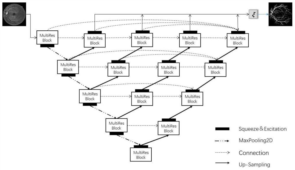
[0033] Embodiment 1: as Figure 1-4 Shown, based on the retinal blood vessel image segmentation method of improving UNet++, the concrete steps of described method are as follows:

[0034] Step1. Randomly crop the retinal images in the DRIVE dataset to expand the dataset;

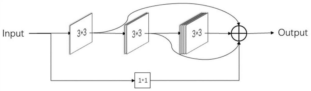
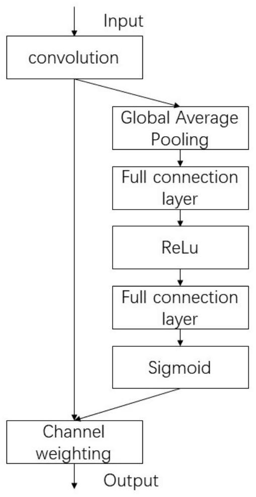
[0035]Step2. Use the MultiRes feature extraction module to extract image features, and use the SeNet module to extract channel attention, and fuse with the image features extracted by the MultiRes feature extraction module to obtain feature maps with different attention weights;

[0036] Step3. Perform the Step2 operation through 4 repetitions, and fuse the features obtained by the 4 Step2 operations through the result of each repetition through a weighted and summed function ξ, and finally use the fused features to segment the retinal blood vessel image;

[0037] Step4. Evaluate the segmentation results of the model by comparing with the manual segmentation results of experts.

[0038] As a preferred solu...


PUM


Abstract

The invention relates to a retinal vessel image segmentation method based on improved UNet + +, and belongs to the technical field of medical image processing. According to the method, a deep supervision network UNet + + is selected as an image segmentation network model, so that the use efficiency of features is improved; a MultiRes feature extraction module is introduced to improve the feature learning effect of small blood vessels in a low-contrast environment, the generalization ability of a network and the expression ability of a network structure are further improved by coordinating features learned by an image in different scales, and a SeNet channel attention module is added to perform extrusion and excitation operation after feature extraction to improve the accuracy of feature extraction of the small blood vessels in the low-contrast environment. Therefore, the receptive field in the feature extraction stage is enhanced, and the weight of a target related feature channel is improved. The improved UNet + + network model is verified based on a DRIVE retina image data set, and compared with an existing model, the evaluation indexes such as the overlapping ratio, the cross-parallel ratio, the accuracy and the sensitivity are improved to a certain extent.

Description

technical field [0001] The invention relates to a retinal vessel image segmentation method based on improved UNet++, in particular to an end-to-end neural network nested retinal vessel image segmentation model method, which belongs to the technical field of medical image processing. Background technique [0002] Fundus retinal vessel image segmentation, as a non-invasive diagnostic method in modern ophthalmology, has become an important part of computer-aided diagnosis of retinal diseases. Such as diabetic retinopathy, hypertension, glaucoma, hemorrhage, venous occlusion and neovascularization and other diseases, regular and accurate measurement of blood vessel width and growth status can provide effective evaluation value for these diseases. Therefore, it is of high application value to conduct vessel segmentation on retinal images to analyze retinal vessel morphology for computer-aided diagnosis of eye diseases. At present, the segmentation of retinal blood vessel images ...

Claims


Application Information

Patent Timeline
Patent Type & Authority Applications(China)
IPC IPC(8): G06T7/00G06T7/10G06N3/04G06N3/08
CPCG06T7/0012G06T7/10G06N3/084G06T2207/20132G06T2207/30041G06N3/048G06N3/045
Inventor 王江峰刘利军冯旭鹏黄青松
Owner KUNMING UNIV OF SCI & TECH
Why Eureka
Try Eureka