Eureka AIR delivers breakthrough ideas for toughest innovation challenges, trusted by R&D personnel around the world.

Intelligent left ventricle magnetic resonance image classification method and device, equipment and medium

A classification method and magnetic resonance technology, applied in the field of medical image processing and image processing, can solve the problems of low accuracy, low efficiency, errors, etc.

Active Publication Date: 2021-05-07
GENERAL HOSPITAL OF PLA +1
View PDF7 Cites 5 Cited by
  • Summary
  • Abstract
  • Description
  • Claims
  • Application Information

AI Technical Summary

Problems solved by technology

[0005] In view of the above-mentioned defects or deficiencies in the prior art, it is desired to provide an intelligent classification method, device, equipment and medium for magnetic resonance images of the left ventricle, which solves the problem of low efficiency due to the need for a large number of manual operations in the entire process in the related art. , introducing errors and low accuracy, making full use of multi-sequence nuclear magnetic imaging data and clinical data to make classification predictions, the results are more reliable and accurate, and by directly using end-to-end classification predictions, no complicated post-processing procedures are required. There is cumulative error, which greatly improves the robustness

Method used


Image

  • Intelligent left ventricle magnetic resonance image classification method and device, equipment and medium
  • Intelligent left ventricle magnetic resonance image classification method and device, equipment and medium
  • Intelligent left ventricle magnetic resonance image classification method and device, equipment and medium

Examples


Embodiment Construction

[0056] The application will be further described in detail below in conjunction with the accompanying drawings and embodiments. It should be understood that the specific embodiments described here are only used to explain related inventions, rather than to limit the invention. It should also be noted that, for ease of description, only parts related to the invention are shown in the drawings.

[0057] It should be noted that, in the case of no conflict, the embodiments in the present application and the features in the embodiments can be combined with each other. The method, device, device and medium for intelligent classification of left ventricular magnetic resonance images according to the embodiments of the present application will be described in detail below with reference to the accompanying drawings and embodiments.

[0058] Before introducing the intelligent classification method for left ventricular magnetic resonance images according to the embodiment of the presen...


PUM


Abstract

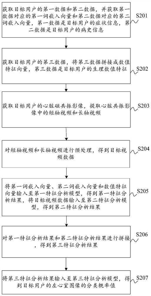
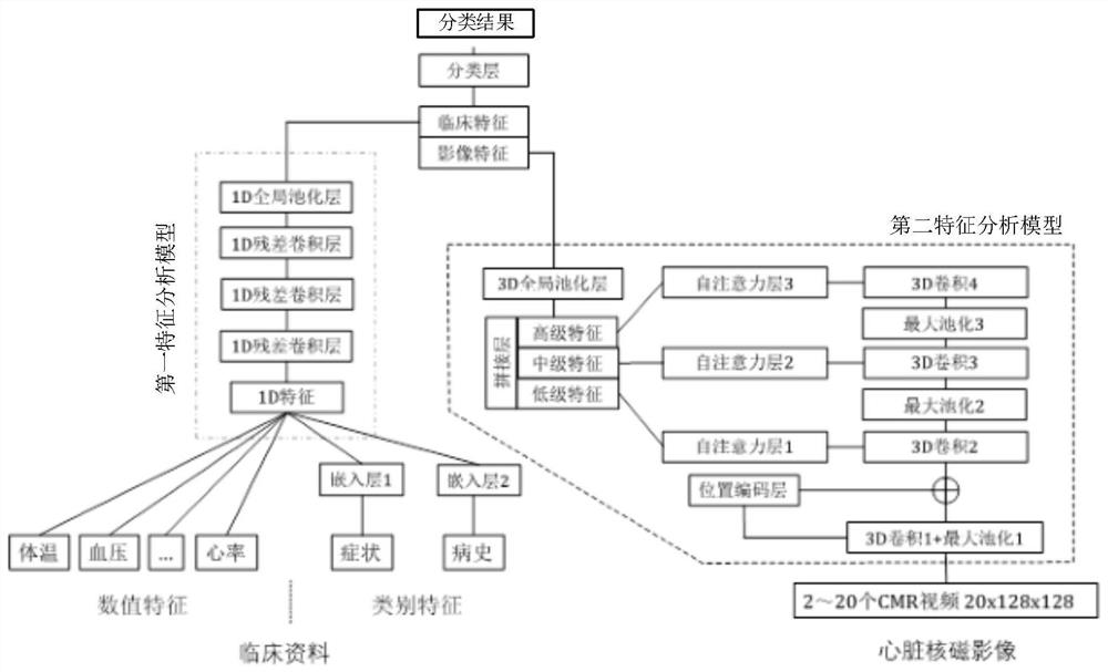
The invention discloses an intelligent left ventricle magnetic resonance image classification method and device, equipment and a medium. The method comprises the steps of obtaining a first word embedding vector corresponding to first data of a detection object, a second word embedding vector corresponding to second data of the detection object, and a numerical feature vector spliced by third data; extracting a short-axis video and a long-axis video in the heart magnetic resonance image of the detected object, and performing preprocessing to obtain target video data; and splicing and inputting the three vectors into the first feature analysis model to obtain a first feature analysis result, inputting the target video data into the second feature analysis model to obtain a second feature analysis result, splicing the first feature analysis result and the second feature analysis result to obtain a third feature analysis result, and inputting the third feature analysis result into the third feature analysis model to obtain a classification probability value of the left ventricle image of the detected object. According to the method, the multi-sequence nuclear magnetic image data and the clinical data are utilized to jointly perform classification prediction, the result is more reliable and accurate, a complex post-processing flow is not needed, accumulated errors do not exist, and the robustness is improved.

Description

technical field [0001] The present disclosure relates to the technical field of image processing, in particular to the field of medical image processing, and in particular to a method, device, device and medium for intelligent classification of magnetic resonance images of the left ventricle. Background technique [0002] With the rapid development of deep learning technology, medical image processing is expected to provide low-cost, efficient and accurate auxiliary medical means. In the field of cardiac diseases, cardiac magnetic resonance has become an important standard for the classification of cardiomyopathy due to its advantages of high resolution, high soft tissue contrast and multi-sequence imaging. [0003] In related technologies, complex processes such as segmentation of MRI images and extraction of cardiac function indicators are usually required. [0004] However, in the whole process, a lot of manual operations are required, resulting in low efficiency, introd...

Claims


Application Information

Patent Timeline
Patent Type & Authority Applications(China)
IPC IPC(8): G06K9/62G06T7/00G06F16/33G06F16/35G06N3/04G06N3/08G16H50/20
CPCG06T7/0012G06F16/35G06F16/3344G06N3/08G16H50/20G06T2207/10088G06T2207/20076G06T2207/20081G06T2207/20084G06T2207/30048G06N3/047G06F18/2415G06F18/241
Inventor 何昆仑李宗任程流泉李瑶张恒王文君张培芳
Owner GENERAL HOSPITAL OF PLA
Why Eureka
Eureka Blog
Learn More