A pressure porous valve chip and its detection method
A chip detection and pressure valve technology, applied in the field of medical devices, can solve problems such as difficulty in operation, achieve the effects of efficient experimentation, strong applicability, and improved detection efficiency
- Summary
- Abstract
- Description
- Claims
- Application Information
AI Technical Summary
Problems solved by technology
Method used
Image
Examples
Embodiment 2
[0052] The method for detecting the chip in embodiment 1 comprises the following steps:
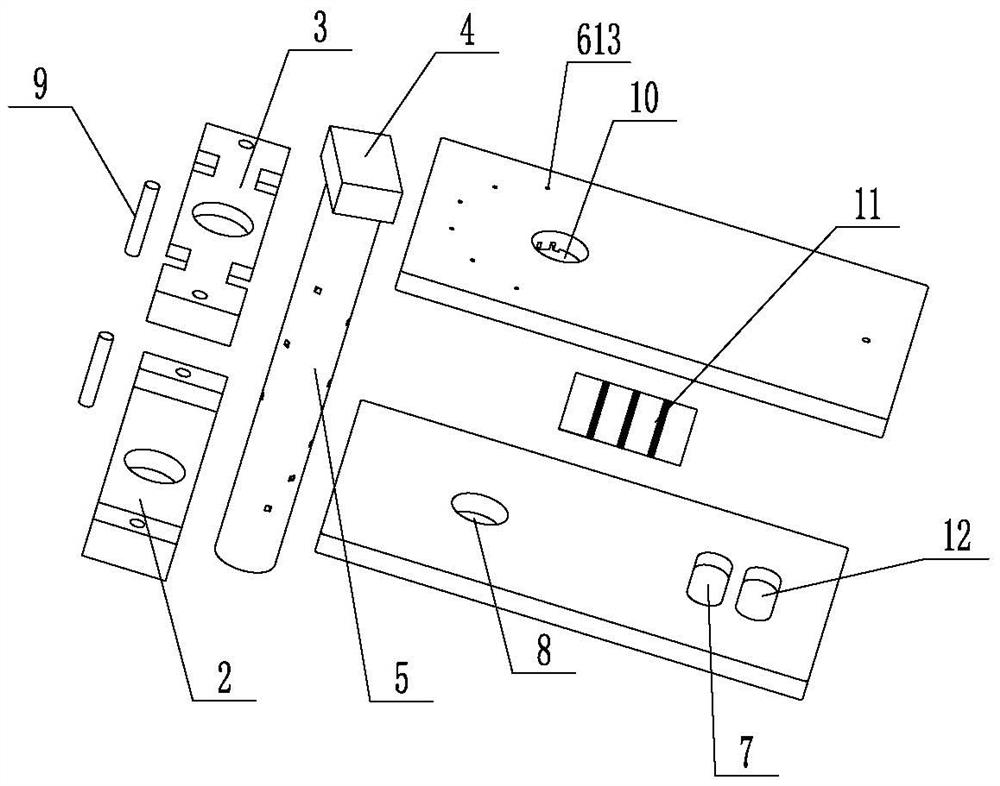
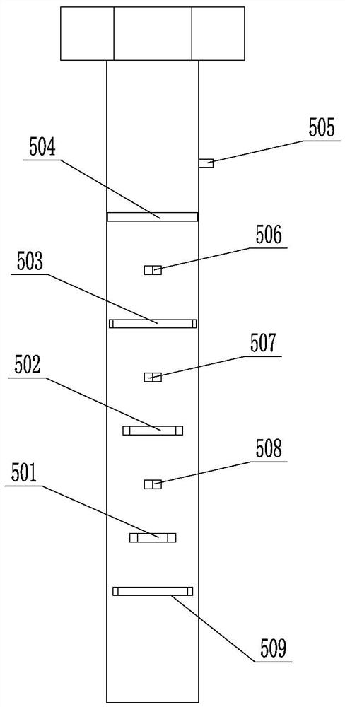
[0053] (1) Add samples to the corresponding liquid storage tanks through each liquid inlet hole 612, put the chip into the supporting detection equipment, the peristaltic pump 14 works, and the samples in the liquid storage tank one 605 are driven by the peristaltic pump 14, Pass through the microfluidic branch channel 613, the fluid through hole 1 509, the flow area 601, and the microfluidic main channel 1 602 to enter the branch reaction channel 611. When the sample starts to enter the buffer tank 7, the peristaltic pump 14 stops and stops for 15 minutes , block the vacant sites of the three capture antibody bands on the antigen-antibody pattern sheet 11 to prevent non-specific adsorption. After 15 minutes of blocking, the peristaltic pump 14 pneumatically sucks out the sample and enters the waste liquid tank 12 or the buffer tank 7. The peristaltic pump 14 stop action;
[0054] (2) Af...
Embodiment 3
[0068] Perform linear detection on the three tumor marker standard products in Example 2, pre-coat 40 μg / mL of AFP capture antibody on the exposed antigen-antibody pattern sheet 11 before detection, use PBS buffer (PH=7.4) as dilution solution, the concentration of AFP standard is 0ng / mL, 5ng / mL, 10ng / mL, 20ng / mL, 40ng / mL, 80ng / mL, 160ng / mL, 320ng / mL, take 30μL and add it to storage tank 2 606 ; Add the 50ug / mL biotin-labeled AFP antibody solution of 30ul in reservoir five; All the other detection steps are the same as in Example 2, and each standard product is measured 3 times with 3 microfluidic chips respectively, and the quantitative analysis system is based on The ratio between the AFP concentration and the gray value of chemiluminescence was fitted to calculate the average value of 8 different concentrations, and a standard curve was drawn.
[0069] Pre-coat the exposed antigen-antibody pattern sheet 11 with 50 μg / mL of CEA capture antibody, use PBS buffer (PH=7.4) as th...
Embodiment 4
[0073] Add the mixed standard sample of AFP and CEA into the liquid storage tank 2 606; add the detection antibody corresponding to the standard in the liquid storage tank 5 609, and the remaining detection steps are the same as in Example 2, and the exposure time of the chemiluminescence instrument is 30s ,get Figure 13-1 image.
[0074] Add the mixed standard sample of CEA and TSGF into the liquid storage tank 2 606; add the detection antibody corresponding to the standard in the liquid storage tank 5, and the remaining detection steps are the same as in Example 2, and the exposure time of the chemiluminescence instrument is 30s. get Figure 13-2 image.
[0075] The mixed standard sample of AFP and TSGF was added to the second storage tank 606; the detection antibody corresponding to the standard was added to the fifth storage tank 609, and the rest of the detection steps were the same as in Example 2. The exposure time of the chemiluminescence instrument was 30s, and ...
PUM
| Property | Measurement | Unit |
|---|---|---|
| linear range | aaaaa | aaaaa |
Abstract
Description
Claims
Application Information
 - R&D
- Intellectual Property
- Life Sciences
- Materials
- Tech Scout
- Unparalleled Data Quality
- Higher Quality Content
- 60% Fewer Hallucinations
Browse by: Latest US Patents, China's latest patents, Technical Efficacy Thesaurus, Application Domain, Technology Topic, Popular Technical Reports.
© 2025 PatSnap. All rights reserved.Legal|Privacy policy|Modern Slavery Act Transparency Statement|Sitemap|About US| Contact US: help@patsnap.com


