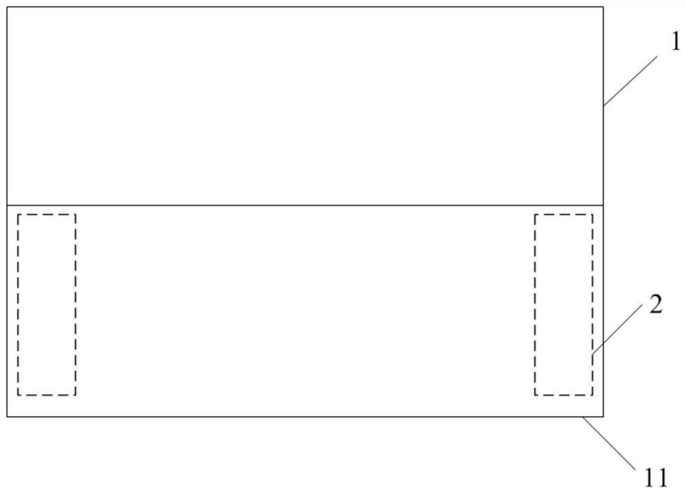
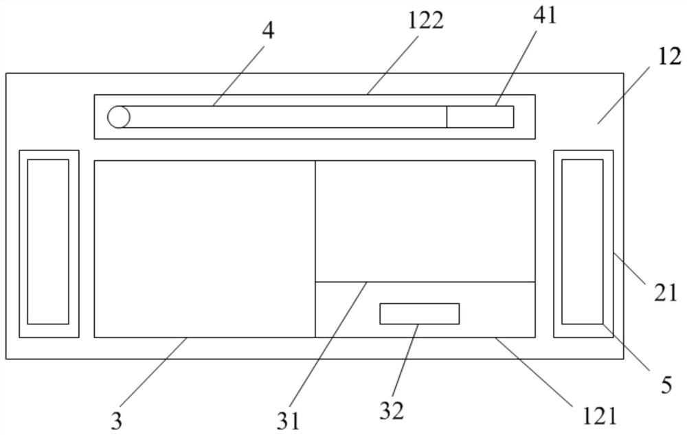
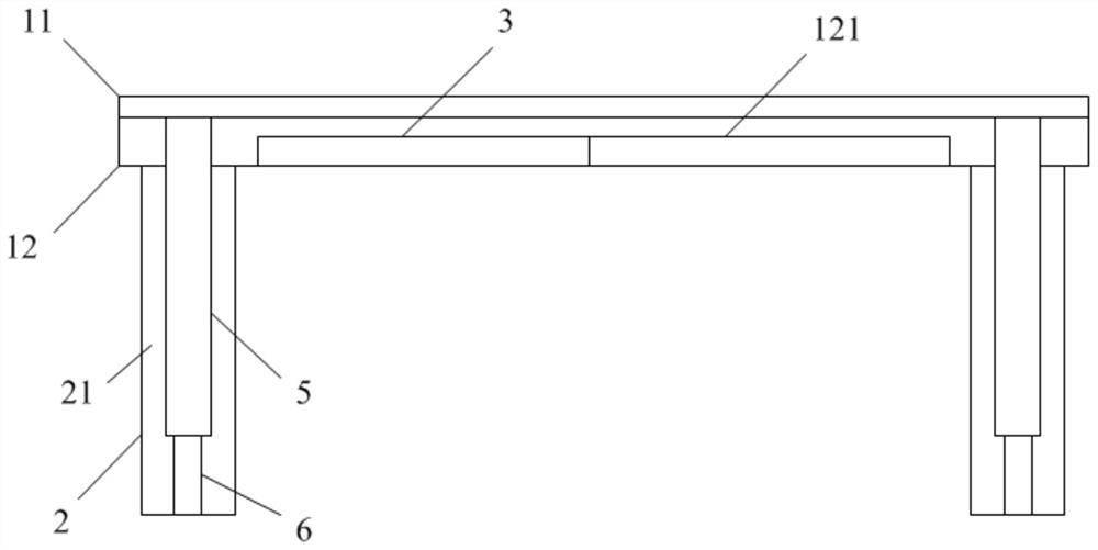
Tumor pathological image display device capable of projecting partitions

A technology for pathological images and display equipment, applied in medical images, legs of general furniture, healthcare informatics, etc., can solve problems such as untargeted enlarged image parts, easy distraction, and doctors unable to watch images for a long time.

Active Publication Date: 2022-06-07
XIAN MEDICAL UNIV
View PDF9 Cites 0 Cited by
  • Summary
  • Abstract
  • Description
  • Claims
  • Application Information

AI Technical Summary

Problems solved by technology

Although the device is convenient for doctors to observe and analyze the image details, the images only stay in front of the doctor repeatedly for a short period of time. Doctors cannot watch the images for a long time and are easily distracted. Moreover, the displayed images of the patented device can only be replaced uniformly. Every doctor The images seen are all the same, and it is impossible to zoom in on different image parts in a targeted manner

Method used


Image

  • Tumor pathological image display device capable of projecting partitions
  • Tumor pathological image display device capable of projecting partitions
  • Tumor pathological image display device capable of projecting partitions

Examples


Embodiment 2

[0044] A tumor pathological image display device capable of projection and separation, the structure is basically the same as that of Embodiment 1, the difference is that in this embodiment, refer to Figure 5 , the projection plate 52 includes a frame 521, and the frame 521 is provided with a plurality of horizontal shading strips 522 arranged in parallel and a plurality of vertical shading strips 523 arranged in parallel. An elliptical cylinder made of rigid opaque materials such as plastic, the two end faces along the length direction are respectively connected to the frame 521 through the rotating device 524. The horizontal shading strips 522 and the longitudinal shading strips 523 have the same structure, and the two end faces of the longitudinal shading strips 523 along the length direction are also rotatably connected to the frame 521 through the rotating device 524 respectively. When the horizontal light-shielding strip 522 and the vertical light-shielding strip 523 ar...

Embodiment 3

[0046] A tumor pathological image display device that can be projected and separated, the structure is basically the same as that of the second embodiment, the difference is that in this embodiment, refer to Figure 6-7, the horizontal shading strip 522 is a cylindrical structure made of a flexible and opaque material such as shading cloth; the rotating device 524 includes a screw 5241, a helical coil spring 5242 sleeved on the screw 5241, and a plurality of nuts 5243. The ends are respectively penetrated and screwed on the side wall of the frame 521 on the same side as the nut 5243 is screwed on the screw 5241 and located on both sides of the side wall of the frame (521), each two nuts 5243 are used to clamp one end of the screw 5241 and the side wall of the frame 521, the central end of the coil spring 5242 is threaded through the screw 5241 and fixed with one end of the extension section at the outer end of the coil spring 5242, and the other end of the extension section is ...

Embodiment 4

[0048] A tumor pathological image display device that can be projected and separated, the structure is basically the same as that of the first embodiment, the difference is that the device of this embodiment also includes a general display screen, and the memory of a U disk or a mobile hard disk can be plugged into the general display screen. , the main display screen is connected with the controller, the main display screen is installed on the wall of the consultation room, and the image displayed on the main display screen is consistent with the image displayed on the fixed display screen 3, which is convenient for the consultation host to guide everyone to watch the image.


PUM


Abstract

The invention belongs to the technical field of auxiliary medical equipment, and in particular relates to a tumor pathological image display device capable of projecting partitions. The equipment includes a controller, a microscope connected to the controller, a memory, and multiple sub-display devices. The sub-display devices include a table top, a vertical board, an image display component, an image capturing component, and a projection component; the table top includes a fixed top panel, a movable top panel The panel and the storage area; the image display component and the image capture component are arranged in the storage area; the storage chamber with the top connected to the storage area is opened in the vertical plate, and the projection assembly is arranged in the storage cavity, and the projection assembly includes a mobile installation boards, lights and projection boards. In the device of the present invention, the image display component is used to display images and partial enlarged images, allowing doctors to watch them for a long time; the image capture component is used to partially capture the displayed images to facilitate observation of image details; Non-destructive division, easy to quickly locate, so that it can adapt to the needs of different doctors.

Description

technical field [0001] The invention belongs to the technical field of auxiliary medical equipment, and in particular relates to a projection-separated tumor pathological image display device. Background technique [0002] Tumors are tissue masses formed by local tissue cell proliferation, and are classified into benign tumors and malignant tumors. Benign tumors grow slowly, are mostly spherical or nodular, do not invade adjacent normal tissues, can be removed by surgery, do not metastasize, and rarely recur. Malignant tumors also become cancers habitually, which have abnormal cell differentiation and proliferation, uncontrolled growth, invasiveness and metastasis, which invade normal tissues and affect the physiological activities of normal organs. [0003] Common methods of tumor diagnosis include imaging observation (X-ray examination, ultrasound examination, CT examination, MRI, etc.), endoscopy (gastroscopy, proctoscopy, colposcopy, etc.) and pathological smear examina...

Claims


Application Information

Patent Timeline
Patent Type & Authority Patents(China)
IPC IPC(8): A47B13/08G16H30/20
CPCA47B13/08G16H30/20
Inventor 王善伟徐文静年申李晟吴庚利聂蕾
Owner XIAN MEDICAL UNIV
Why Eureka
Try Eureka