Graphical longitudinal display for intraluminal ultrasound imaging and associated devices, systems, and methods

An ultrasonic imaging system and ultrasonic imaging technology, applied in ultrasonic/sonic/infrasonic Permian technology, ultrasonic/sonic/infrasonic image/data processing, ultrasonic/sonic/infrasonic diagnosis, etc., can solve problems such as time required

Pending Publication Date: 2021-06-04
KONINKLJIJKE PHILIPS NV +1
View PDF10 Cites 1 Cited by
  • Summary
  • Abstract
  • Description
  • Claims
  • Application Information

AI Technical Summary

Problems solved by technology

This calculation takes time and may require interaction with sterile equipment

Method used


Image

  • Graphical longitudinal display for intraluminal ultrasound imaging and associated devices, systems, and methods
  • Graphical longitudinal display for intraluminal ultrasound imaging and associated devices, systems, and methods
  • Graphical longitudinal display for intraluminal ultrasound imaging and associated devices, systems, and methods

Examples


Embodiment Construction

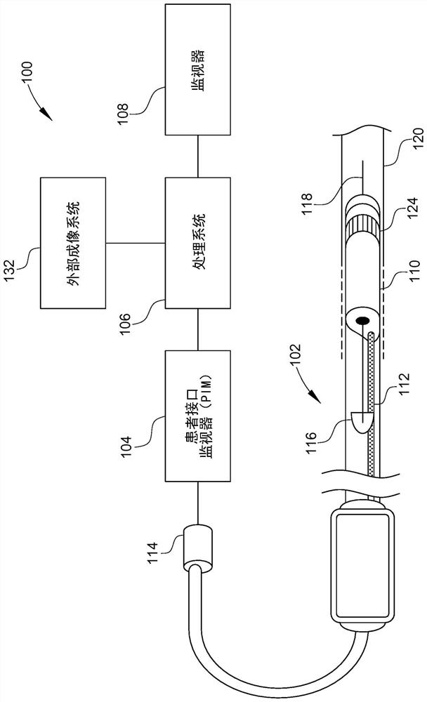
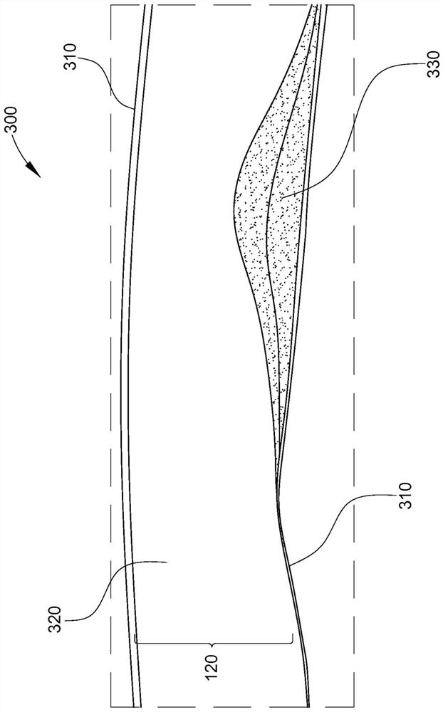
[0031] The present disclosure relates generally to medical imaging, including imaging in association with a lumen of a patient's body using an intraluminal imaging device. For example, the present disclosure describes systems, devices, and methods for visually summarizing images, measurements, and calculations acquired during an IVUS pullback procedure or other intravascular procedure. In accordance with at least one embodiment of the present disclosure, there is provided a system for representing a plurality of lumens in a longitudinal format using a graphical representation indicating the diameter, area, percent compression, or percent improvement of the lumen at each location along retraction. tomographic image. This allows clinicians and other users to view a single screen display that includes clinically relevant data from dozens or hundreds of intraluminal ultrasound images in a visually simplified format. This system is hereinafter referred to as a graphics portrait di...


PUM


Abstract

Disclosed is an intraluminal ultrasound imaging system, including a processor circuit in communication with an intraluminal ultrasound imaging catheter, and configured to receive a plurality of intraluminal ultrasound images obtained by the imaging catheter while the imaging catheter is moved through a body lumen of a patient, wherein the plurality of intraluminal ultrasound images comprise axial cross-sectional views of the body lumen. The processor circuit is further configured to generate a stylized graphic of the body lumen based on the plurality of intraluminal ultrasound images, wherein the stylized graphic comprises a longitudinal cross- sectional view of the body lumen, and output, to a display in communication with the processor circuit, a screen display comprising the stylized graphic.

Description

[0001] Cross References to Related Applications [0002] This application claims priority and benefit to US Provisional Patent Application US62 / 751,289 filed October 26, 2018, the entire contents of which are hereby incorporated by reference. technical field [0003] The subject matter described herein relates to systems for medical imaging. Specifically, the disclosed system provides a graphical longitudinal display in a simplified visual format to facilitate the acquisition and interpretation of peripheral intravascular ultrasound or IVUS images during the withdrawal procedure. The system has particular, but not exclusive, utility for the diagnosis and treatment of vascular disease. Background technique [0004] Plaques such as angioplasty and stenting in peripheral veins (inferior vena cava - IVC, ilia, femoral vein), IVC filter retrieval, EVAR and FEVAR (and similar abdominal features) Peripheral vascular surgery with resection and thrombectomy is a procedure using IVU...

Claims


Application Information

Patent Timeline
Patent Type & Authority Applications(China)
IPC IPC(8): A61B8/12A61B8/00A61B8/08
CPCA61B8/12A61B8/461A61B8/5223A61B8/0841A61B8/5207A61B8/5215A61B8/523
Inventor 赵沛然A·奈尔N·S·拉伊古鲁Y·吉利斯
Owner KONINKLJIJKE PHILIPS NV
Why Eureka
Try Eureka