Wearable bladder urine volume ultrasonic monitoring system

An ultrasonic monitoring and wearable technology, applied in the clinical medical field, can solve the problems of complex imaging system algorithm, high requirements on placement accuracy, low accuracy, etc., to overcome low spatial resolution, easy real-time measurement, and time-consuming measurement. less effect

Active Publication Date: 2021-06-15
SUZHOU INST OF BIOMEDICAL ENG & TECH CHINESE ACADEMY OF SCI
View PDF10 Cites 2 Cited by
  • Summary
  • Abstract
  • Description
  • Claims
  • Application Information

AI Technical Summary

Problems solved by technology

But its main disadvantages are: 1. Single-probe measurement has low spatial resolution and low accuracy; 2. Due to the need to detect the position of the small intestine behind the bladder, the accuracy of its placement is high, and third-party equipment and doctor assistance are required.
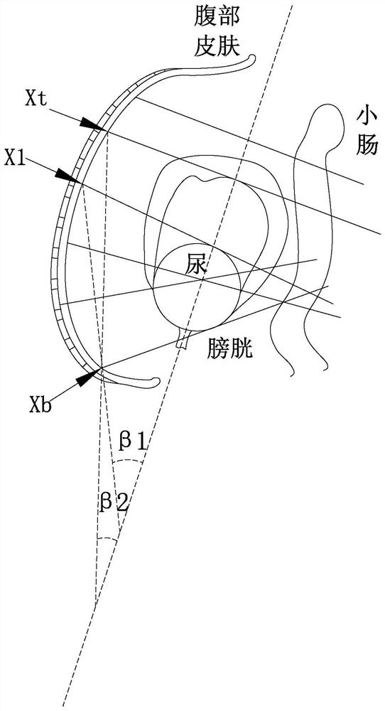
[0012]Therefore, the use of ultrasound to monitor bladder urine volume mainly uses a single-array element probe for detection, and the imaging of a single-array element ultrasonic probe takes 1-2 minutes, and the space The resolution is low, and there is a large error rate in the measurement results; if a 3D image is required to scan the bladder, the imaging system algorithm is complex, the measurement time is long, the equipment is expensive, and the instrument is bulky, which is not suitable for real-time measurement

Method used


Image

  • Wearable bladder urine volume ultrasonic monitoring system
  • Wearable bladder urine volume ultrasonic monitoring system
  • Wearable bladder urine volume ultrasonic monitoring system

Examples


Embodiment Construction

[0045]In order to make the above objects, features and advantages of the present invention more obvious and understandable, the present invention will be described in detail below in conjunction with the accompanying drawings and specific embodiments. In the following description, numerous specific details are set forth in order to provide a thorough understanding of the present invention. However, the present invention can be implemented in many other ways different from those described here, and those skilled in the art can make similar improvements without departing from the connotation of the present invention, so the present invention is not limited by the specific embodiments disclosed below.

[0046] In describing the present invention, it should be understood that the terms "center", "longitudinal", "transverse", "length", "width", "thickness", "upper", "lower", "front", " Back", "Left", "Right", "Vertical", "Horizontal", "Top", "Bottom", "Inner", "Outer", "Clockwise",...


PUM


Abstract

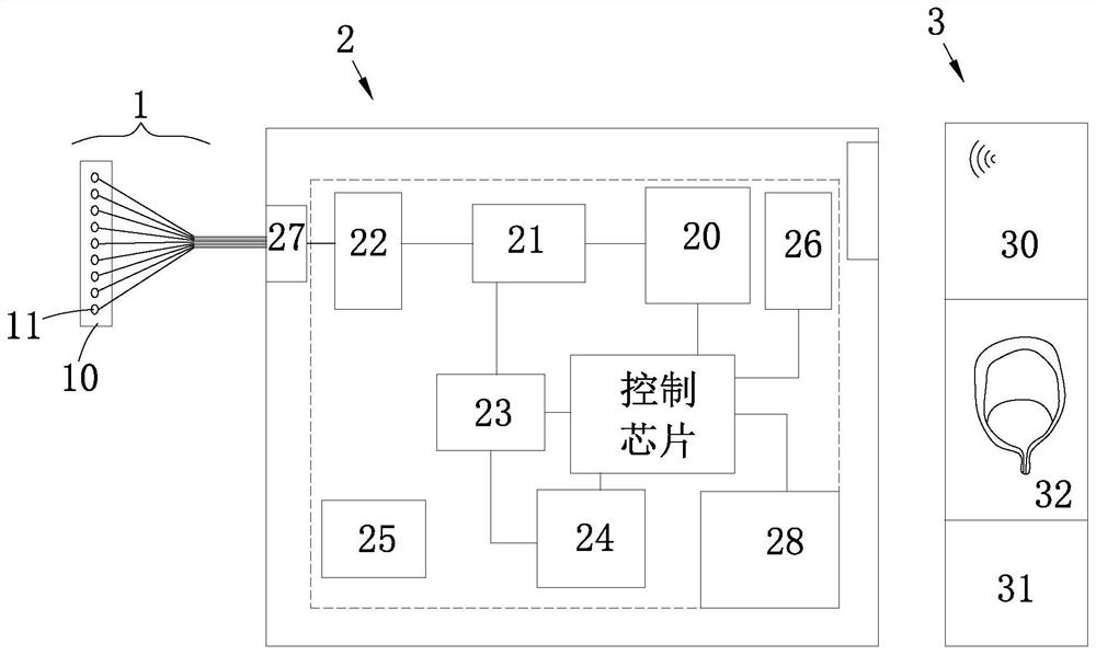
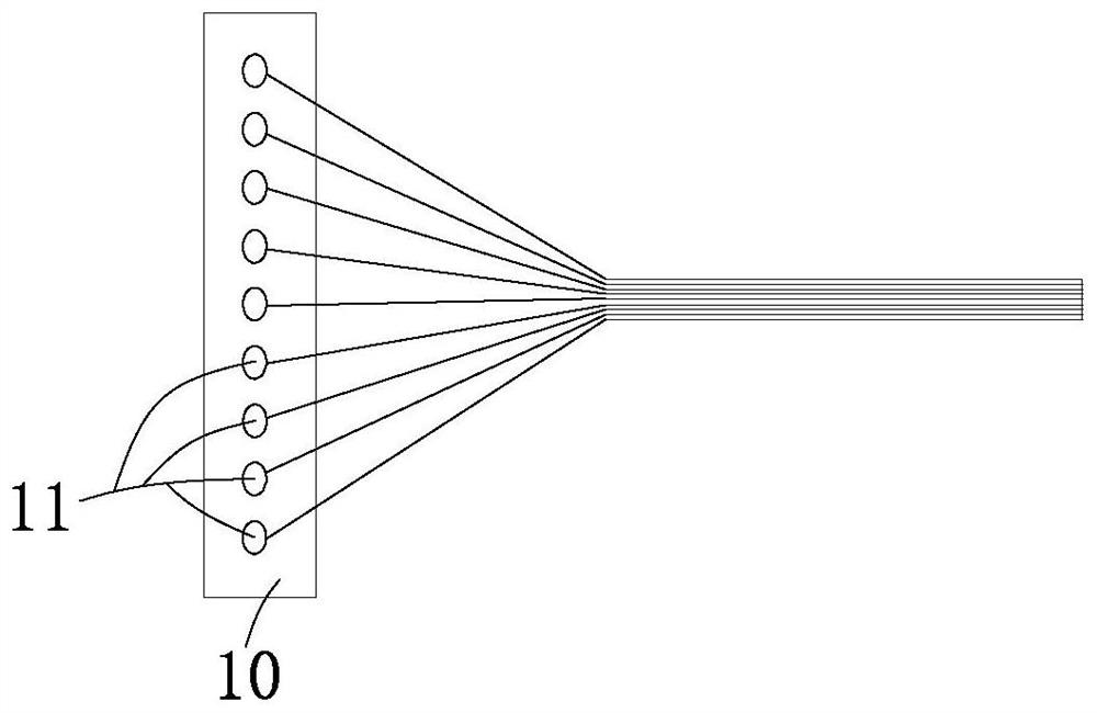
The invention relates to a wearable bladder urine volume ultrasonic monitoring system, which comprises an ultrasonic detection unit, a host and a system terminal, the ultrasonic detection unit comprises a flexible stretchable wearable flexible substrate and a plurality of ultrasonic probes distributed on the flexible substrate, the host comprises a high-voltage excitation emitter, a T / R switch, a gating circuit controller, an AFE receiver, a data processor, a wireless transmission module and a power supply module, and the system terminal comprises a data receiving module, a data analysis module and a post-processing display module. On one hand, the bladder volume can be calculated without bladder imaging or reexamination, the defects that an existing single-array-element sensor is low in spatial resolution and poor in accuracy, needs professional auxiliary operation and the like can be overcome, the state of urine in the bladder can be conveniently obtained, and corresponding auxiliary information is provided for clinical monitoring and urination of a patient; and on the other hand, wearing is convenient, real-time measurement is facilitated, measurement time consumption is low, manufacturing cost is low, and the size is small.

Description

technical field [0001] The invention belongs to the technical field of clinical medicine, and in particular relates to a wearable ultrasonic monitoring system for bladder urine volume. Background technique [0002] Bladder residual urine volume is often used as an important basis for understanding bladder function and judging urethral obstruction. Online monitoring is very important. [0003] However, at present, the residual urine volume of the bladder is mainly judged according to the size of the bladder volume, and the research hotspots of bladder volume measurement methods mainly include implanted sensor method, bioimpedance method and in vitro imaging method. [0004] Specifically, the implanted sensor method: the implantable strain sensor is implanted in the body, and the signal emitted can be detected by an external receiver, and its frequency corresponds to the measured pressure. There is a linear relationship between bladder volume and pressure. However, this app...

Claims


Application Information

Patent Timeline
Patent Type & Authority Applications(China)
IPC IPC(8): A61B8/00
CPCA61B8/4444A61B8/4427A61B8/4488A61B8/4236
Inventor 简小华徐杰
Owner SUZHOU INST OF BIOMEDICAL ENG & TECH CHINESE ACADEMY OF SCI
Why Eureka
Try Eureka