
Image feature extraction method based on deep learning

A technology of deep learning and feature extraction, applied in the field of medical image processing, can solve problems such as overfitting, difficulty in ensuring time efficiency, and time-consuming, etc., and achieve the effect of fast positioning

Active Publication Date: 2021-08-10
广州天鹏计算机科技有限公司
View PDF9 Cites 0 Cited by
  • Summary
  • Abstract
  • Description
  • Claims
  • Application Information

AI Technical Summary

Problems solved by technology

For the deep learning method, although it has been well applied in the medical image classification session, there are many parameters, so it is easy to cause the problem of overfitting. It takes a lot of time to train the deep learning model, and it is difficult to guarantee the current large-scale Time Efficiency in a Data Environment

Method used


Image

  • Image feature extraction method based on deep learning

Examples


Embodiment Construction

[0027] The following and accompanying appendices illustrating the principles of the invention Figure 1 A detailed description of one or more embodiments of the invention is provided together. The invention is described in connection with such embodiments, but the invention is not limited to any embodiment. The scope of the invention is limited only by the claims and the invention encompasses numerous alternatives, modifications and equivalents. In the following description, numerous specific details are set forth in order to provide a thorough understanding of the present invention. These details are provided for the purpose of example and the invention may be practiced according to the claims without some or all of these specific details.

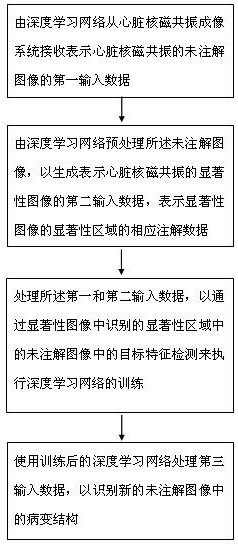
[0028] One aspect of the present invention provides an image feature extraction method based on deep learning. figure 1 It is a flow chart of an image feature extraction method based on deep learning according to an embodiment of the p...


PUM


Abstract

The invention provides an image feature extraction method based on deep learning. The method comprises the following steps: receiving first input data representing an unannotated image of heart nuclear magnetic resonance from a heart nuclear magnetic resonance imaging system by a deep learning network; pre-processing the unannotated image by a deep learning network to generate second input data representing a saliency image of cardiac nuclear magnetic resonance and corresponding annotation data representing a saliency region of the saliency image; processing the first and second input data to perform training of a deep learning network by target feature detection in an unannotated image in a saliency region identified in a saliency image; and processing the third input data by using the trained deep learning network to identify a lesion structure in a new unannotated image. According to the image feature extraction method based on deep learning, a complex structure is not needed, training of a needed model is completed at a time, and heart disease variable structure positioning is carried out more quickly.

Description

technical field [0001] The invention relates to medical image processing, in particular to an image feature extraction method based on deep learning. Background technique [0002] Machine learning-based methods are able to automatically classify individuals. Corresponding to cardiac MRI images, there are characteristics such as small sample size and high feature dimension. Traditional machine learning methods only use simple shallow classification models, which cannot fully mine the advanced features of cardiac functional data. At the same time, these methods cannot effectively exploit the topological information in fNMR data. Therefore, the classification effect of fMRI data based on traditional machine learning methods needs to be further improved. For the deep learning method, although it has been well applied in the medical image classification session, there are many parameters, so it is easy to cause the problem of overfitting. It takes a lot of time to train the dee...

Claims


Application Information

Patent Timeline
IPC IPC(8): G06T7/00G06T7/90G06K9/46G06N3/04G06N3/08
CPCG06T7/0012G06T7/90G06N3/08G06T2207/10088G06T2207/20081G06T2207/20104G06T2207/30048G06V10/44G06V10/56G06N3/045
Inventor 叶方全
Owner 广州天鹏计算机科技有限公司
Learn More