Blood vessel dilation device and method

A blood vessel expansion and blood vessel technology, applied in the field of medical devices and its clinical application, can solve the time-consuming and labor-intensive problems of micro-vascular anastomosis and suture process, and achieve the effect of reducing the chance of re-embolization, reducing the difficulty of blood vessel suturing, and improving the speed of blood vessel repair

Inactive Publication Date: 2021-08-31
徐世保
View PDF0 Cites 0 Cited by
  • Summary
  • Abstract
  • Description
  • Claims
  • Application Information

AI Technical Summary

Problems solved by technology

[0004] The present invention improves the time-consuming and labor-intensive problems in the microvascular anastomosis and suturing process in the prior art, and provides a reasonable structure, efficient and reliable vascular expansion device and its method

Method used


Image

  • Blood vessel dilation device and method
  • Blood vessel dilation device and method
  • Blood vessel dilation device and method

Examples


Embodiment Construction

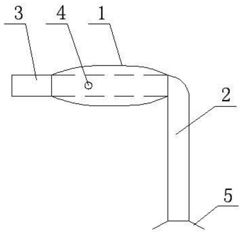
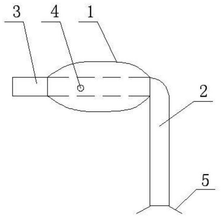
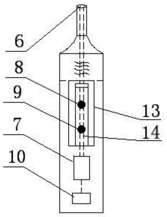
[0027] The vasodilation apparatus and method of the present invention will be further described below in conjunction with the accompanying drawings and specific embodiments: Connect the syringe interface 6 of the automatic charge and discharge device 6, the absorbable blood stent is attached to the outside of the balloon dilator, where the balloon dilator contains balloon 1, L-shaped metal catheter 2 and blunt clip 3, blunt The end of the end of the linear needle 3 is connected to the first end of the L-shaped metal conduit 2, and the middle portion of the L-shaped metal conduit 2 on the first end side is provided with a side vent hole 4, and the end side of the L-shaped metal conduit 2 is connected to the end side. Port 5, can be connected to the pen automatic charge and discharge device or syringe.

[0028] The balloon 1 is designed such as a tension, which is blue, which is convenient for distraction of the absorbable bracket to distinguish the blood tube wall. The diameter of ...


PUM

PropertyMeasurementUnit
Lengthaaaaaaaaaa

Abstract

The invention discloses a blood vessel dilation device and method. The problem that time and labor are wasted in the micro blood vessel anastomosis and suturing process in the prior art is solved. The blood vessel dilation device comprises a balloon dilator, an automatic liquid charging and discharging device and an absorbable blood stent, the balloon dilator is connected with an injector connector of the automatic liquid charging and discharging device, and the absorbable blood stent sleeves the outer part of the balloon dilator; and the balloon dilator comprises a balloon, an L-shaped metal catheter and a blunt straight needle, the tail end of the blunt straight needle is connected with the head end of the L-shaped metal catheter, a lateral vent hole is formed in the middle of the L-shaped metal catheter on the head end side, and a connector is arranged at the tail end of the L-shaped metal catheter on the tail end side. The technology is applied to clinical surgical medical treatment, a good auxiliary method is provided for pure blood vessel anastomosis, the blood vessel suture difficulty can be reduced, the blood vessel repair speed can be increased, and the probability of postoperative reembolism can be reduced by placing the blood vessel stent.

Description

Technical field [0001] The present invention relates to medical equipment and its clinical applications, and in particular, to a vascular expansion device and a method thereof. Background technique [0002] For the anastomosis of the vasodilator, such as the retraction, vein repair, due to the thickness of the lumen, the front and rear wall of the lumen can be clearly separated, the naked eye or the auxiliary magnifier can be sewed. However, for the repair of medium and small blood vessels, especially the veins of the arteries, the front and rear walls are easy to fit, causing the stitching difficulties, and difficult to control the stitching of the non-full sewing. There is happening. In order to change this situation, currently common methods are magnetized, anastomotic, needle ring method, and artificial blood huger kiss. Magnetic pipe method can quickly suck together, reduce the sewing process, but there is no magnetic force, large volume, and need to be in vivo in vivo. The ...

Claims


Application Information

Patent Timeline
IPC IPC(8): A61B17/00A61B17/02A61F2/88A61F2/958A61M25/10A61M29/04
CPCA61B17/00A61B17/02A61B2017/00004A61B2017/00778A61F2/88A61F2/958A61M25/10A61M29/02A61M2210/12A61M2210/1425A61M2210/163A61M25/10182A61M2210/005
Inventor 徐世保
Owner 徐世保
Why Eureka
Try Eureka