Medical image segmentation model compression method

A technology for segmenting models and medical images, applied in the field of medical image processing, can solve problems such as difficult to achieve sub-networks and small sub-networks, and achieve the effects of reducing computing costs, reducing computing costs, and optimizing model structure.

Active Publication Date: 2021-09-17
BEIJING UNIV OF POSTS & TELECOMM
View PDF7 Cites 2 Cited by
  • Summary
  • Abstract
  • Description
  • Claims
  • Application Information

AI Technical Summary

Problems solved by technology

However, since the size of the sub-network is smaller than that of the base network, it is difficult to train the sub-network from scratch to achieve comparable results to the base network.

Method used


Image

  • Medical image segmentation model compression method
  • Medical image segmentation model compression method
  • Medical image segmentation model compression method

Examples


Embodiment Construction

[0034] In order to better understand the technical solution, the method of the present invention will be described in detail below in conjunction with the accompanying drawings.

[0035] The invention provides a method for compressing a medical image segmentation model, comprising the following steps:

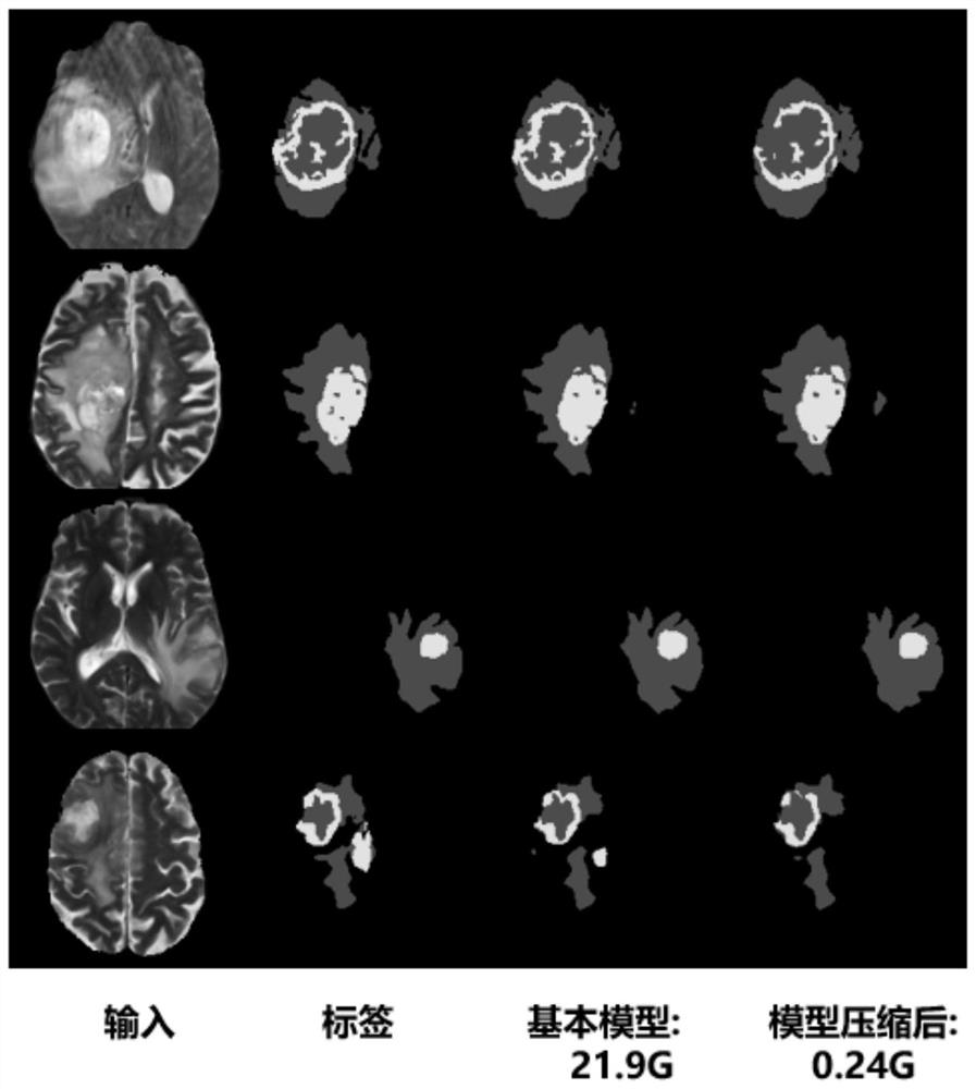
[0036] Step S1, collect the data in the medical image database. In this embodiment, the magnetic resonance images of brain tumor patients are taken as an example, mainly including four modalities: T1, T1c, T2 and FLAIR.

[0037] Step S2, perform data preprocessing, including motion correction, spatial standardization, grayscale normalization, scalp and neck removal, and size cropping. Then the 3D MRI of each subject was centrally cropped, the entire brain area was reserved, and the black area of ​​​​the border was removed.

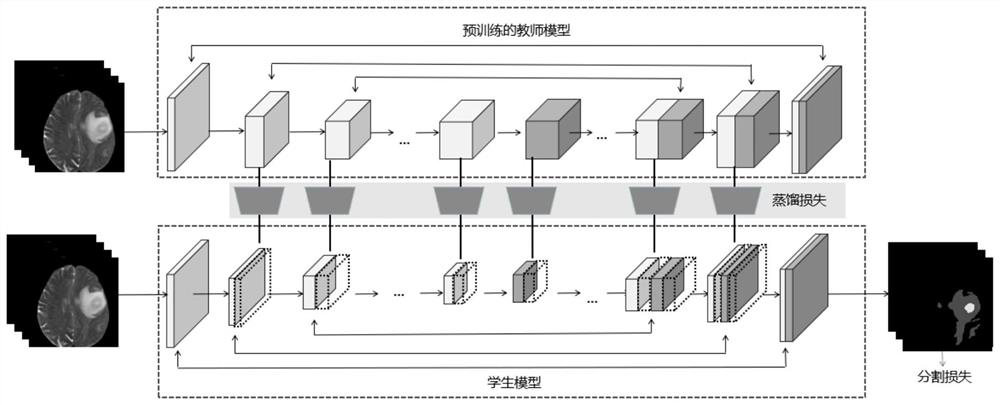
[0038] In step S3, Res-Unet is used as the basic skeleton of the network, and separable convolution is used as the convolutional layer. Res-Unet is a res...


PUM


Abstract

The invention discloses a medical image segmentation model compression method, and belongs to the field of medical image processing. The method comprises the following steps: aiming at a medical image segmentation basic model, constructing a search space according to the number of convolution kernels used at each position in the model, aiming at a coding-decoding structure of a segmentation network, searching a sub-network with small calculation amount and high segmentation precision in the search space by using a symmetric neural network, and carrying out segmentation on the sub-network, wherein the coding and decoding structures are symmetrical, and weight sharing policies are used to mitigate computational costs and training resources when traversing the entire search space; finally, by using a knowledge distillation method in the network training process, the basic model serving as a teacher mode, the compressed sub-network serving as a student model, and completing the knowledge transfer between the basic model and the student model. Through neural network search and knowledge distillation, the calculation cost of network construction is greatly reduced on the premise of ensuring the segmentation effect of the medical image segmentation model, and the method can be applied to various medical image segmentation models.

Description

technical field [0001] The invention relates to the field of medical image processing, in particular to a method for compressing medical image segmentation models based on neural network search and knowledge distillation. Background technique [0002] The task of medical image segmentation has been a research hotspot in the fields of computer vision and nature. With the rapid development and application of Convolutional Neural Networks (CNNs), more and more medical segmentation models based on Deep Learning (DL) have been proposed, and have achieved good results in many disease segmentation tasks. results. On the one hand, the number of layers of neural networks is increasing; on the other hand, the development of medical equipment also provides higher resolution data. This makes the medical image segmentation task better and better, but the size of the model is getting bigger and bigger, which is not convenient for the application of the model and the deployment on the ha...

Claims


Application Information

Patent Timeline
Patent Type & Authority Applications(China)
IPC IPC(8): G06T7/00G06T7/11G06N3/04G06N3/08
CPCG06T7/0012G06T7/11G06N3/08G06T2207/20081G06T2207/30004G06N3/045
Inventor 康桂霞胡凤明郑重
Owner BEIJING UNIV OF POSTS & TELECOMM
Why Eureka
Try Eureka