
Medical image detection equipment with anti-collision and anti-shaking functions

A technology of medical imaging and detection equipment, applied in the field of gynecological examination, can solve problems such as easy influence on the surrounding environment, lower detection safety, and damage to the inner wall of the vagina, and achieve the effects of improving the accuracy of judgment, reducing shaking, and not easy to fall off

Pending Publication Date: 2021-10-19
卢玉蓉
View PDF1 Cites 2 Cited by
  • Summary
  • Abstract
  • Description
  • Claims
  • Application Information

AI Technical Summary

Problems solved by technology

[0006] Aiming at the problems in the prior art, the object of the present invention is to provide a medical image detection device with anti-collision and anti-shake functions, which solves the problem that the traditional detection devices are all passed through the female vagina. into the female vagina, and manually turn the vaginal detection device inserted into the female vagina to observe the inner wall of the vagina. If the female is lying on the transfer bed for detection, the hands of the medical staff must always be inserted into the reproductive position of the female student. It is easy to operate and inconvenient during the transfer process, and it is easy to affect the surrounding environment. In addition, during the transfer process, medical staff need to frequently turn the vagina detection device inserted into the female vagina to achieve multi-angle observation of the vaginal wall. It is easy to shake, so that the vaginal detection device inserted into the female vagina while rotating during the transfer process is likely to cause damage to the inner wall of the vagina and reduce the technical problem of detection safety

Method used


Image

  • Medical image detection equipment with anti-collision and anti-shaking functions
  • Medical image detection equipment with anti-collision and anti-shaking functions
  • Medical image detection equipment with anti-collision and anti-shaking functions

Examples


Embodiment Construction

[0032] In order to make the technical means, creative features, goals and effects achieved by the present invention easy to understand, the present invention will be further elaborated below in conjunction with specific drawings. It should be noted that, in the case of no conflict, the embodiments and Features in the embodiments can be combined with each other.

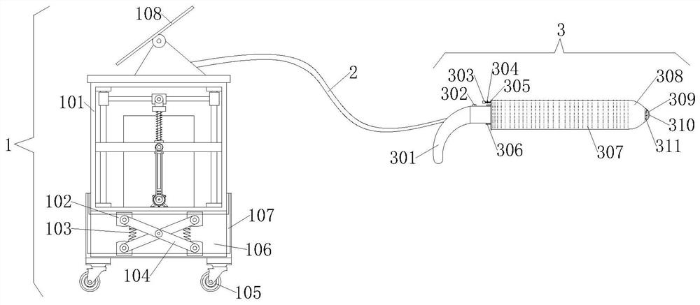
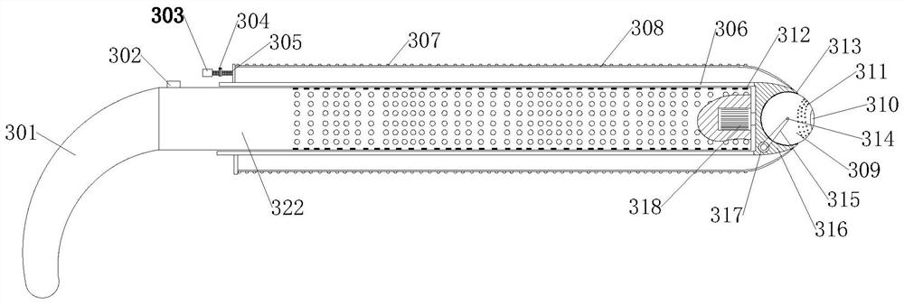
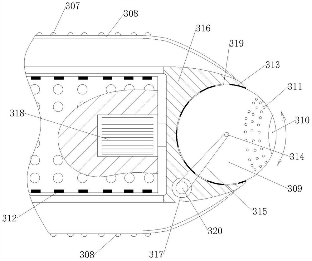
[0033] see Figure 1-5 It is a schematic diagram of the overall structure of a medical image detection device with anti-collision and anti-shake functions;

[0034] A medical image detection device with anti-collision and anti-shake functions, which is applied in the vagina of a woman who is about to give birth, and performs real-time monitoring during the body transfer process of a woman who is about to give birth, including display and observation equipment 1, communication cable 2, and vagina detection Stick 3, the vagina detection stick 3 is inserted into the vagina of a woman who is about to give birth, and the ...


PUM


Abstract

The invention provides medical image detection equipment with an anti-collision and anti-shake function, and relates to the technical field of gynecologic examination, the medical image detection equipment comprises display observation equipment, a communication cable and a vagina detection rod, the vagina detection rod extends into the vagina of a woman to be delivered, the display observation device is placed on the ground or a transferred vehicle, the vagina detection rod transmits data to the display and observation equipment in real time through a communication cable for processing and analysis, the display and observation equipment comprises a main case and a bottom supporting seat arranged below the main case, and calculation parts for processing the data transmitted by the communication cable are stored in the main case; the multiple second cameras are arranged on the outer circle face of the detection rod at equal intervals, the vagina wall is vertically irradiated through the second cameras on the outer circle face of the detection rod, medical staff can more clearly observe the contraction condition of the vagina inner wall of a woman about to be delivered through the display panel, and the judgment accuracy of the medical staff is improved.

Description

technical field [0001] The invention relates to the technical field of gynecological examination, in particular to a medical image detection device with anti-collision and anti-shake functions. Background technique [0002] Peripartum female friends should be admitted to the hospital in time to prepare for delivery when they have labor symptoms. After arriving at the hospital, the doctor will arrange female friends to prepare for delivery in the delivery room. Sometimes, for perinatal female friends who are about to give birth due to family If the distance to the hospital is relatively far or the scale of the hospital is relatively large, the doctor will first arrange for the female friend to be transferred to the waiting room. During the transfer process, due to the transportation, it takes a long time, and real-time monitoring of the vagina and cervix of the woman is required during the transfer process. To determine the changing characteristics of the vagina and uterine o...

Claims


Application Information

Patent Timeline
IPC IPC(8): A61B1/303A61B1/002A61B1/04A61B1/06
CPCA61B1/303A61B1/06A61B1/04A61B1/002A61B1/00147A61B1/00071
Inventor 卢玉蓉
Owner 卢玉蓉
Learn More