
Medical image automatic labeling method and system under small sample condition

A medical image and automatic labeling technology, applied in the field of image processing, can solve problems such as laborious, time-consuming, and manual labeling method errors, and achieve good clinical significance, sample balance between classes, and rapid generation effects

Pending Publication Date: 2021-11-02
AFFILIATED ZHONGSHAN HOSPITAL OF DALIAN UNIV
View PDF0 Cites 0 Cited by
  • Summary
  • Abstract
  • Description
  • Claims
  • Application Information

AI Technical Summary

Problems solved by technology

[0005] The shape of the lesions in the image is often irregular, and manual labeling takes a lot of time. The automatic labeling method learned by artificial intelligence often has problems in accuracy, and it cannot guarantee that every image is correctly labeled. Using the above methods alone The time to form a certain number of standard data sets cannot be shortened
Therefore, the existing technology has technical defects such as time-consuming and laborious, and cannot quickly generate a standard labeling data set; moreover, the manual labeling method is easy to form wrong labels due to subjective factors, and the labeling effect of the manual revision method under pre-labeling is poor. Ensure that each image is correctly labeled, and thus cannot guarantee the correctness of the standard labeled dataset

Method used


Image

  • Medical image automatic labeling method and system under small sample condition

Examples


Embodiment Construction

[0034] The following will clearly and completely describe the technical solutions in the embodiments of the present invention with reference to the accompanying drawings in the embodiments of the present invention. Obviously, the described embodiments are only some, not all, embodiments of the present invention. Based on the embodiments of the present invention, all other embodiments obtained by persons of ordinary skill in the art without making creative efforts belong to the protection scope of the present invention.

[0035] In order to make the above objects, features and advantages of the present invention more comprehensible, the present invention will be further described in detail below in conjunction with the accompanying drawings and specific embodiments.

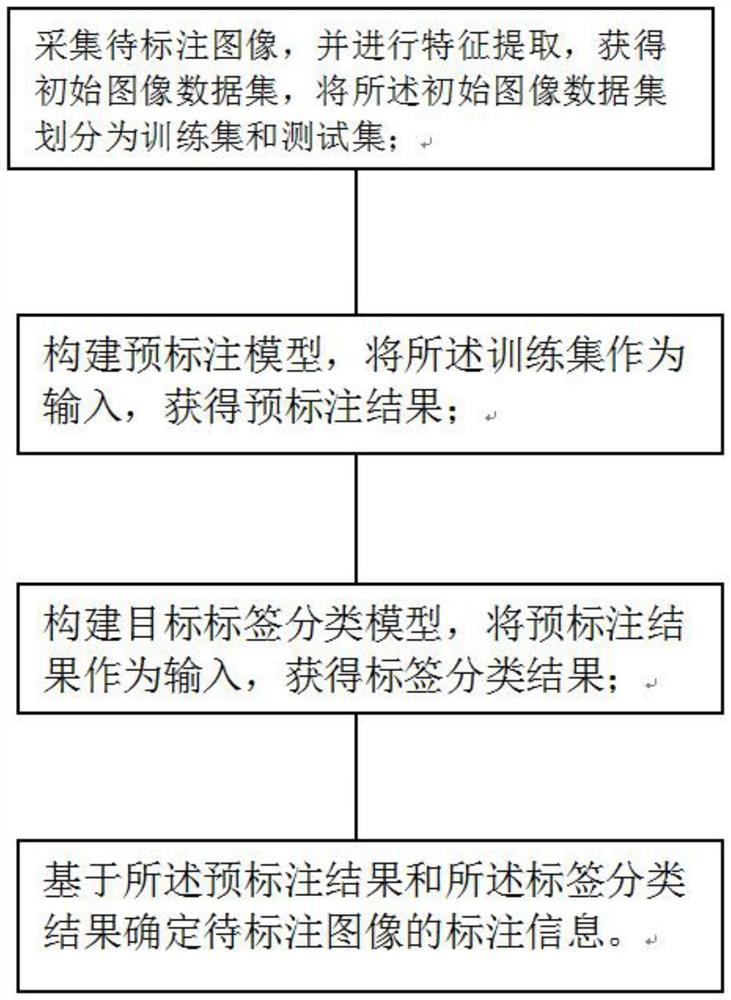
[0036] Such as figure 1 As shown, the present invention provides a method and system for automatic labeling of medical images under small sample conditions, wherein the method specifically includes the following s...


PUM


Abstract

The invention discloses a medical image automatic labeling method and system under a small sample condition, and the system comprises an image collection module, a labeling processing module, a label classification module and a labeling generation module which are connected in sequence. The method comprises: through collecting a to-be-labeled image and carrying out the feature extraction of the to-be-labeled image, obtaining an initial image data set; dividing the initial image data set into a training set and a test set in proportion; constructing a pre-labeling model, and taking the training set as the input of the pre-labeling model to obtain a pre-labeling result; constructing a target label classification model, and taking the pre-labeling result as the input of the label classification model to obtain a label classification result; and determining annotation information of the to-be-annotated image based on the pre-annotation result and the tag classification result. According to the method and the system, automatic intelligent labeling of the medical image is realized, manual outline sketching of the image, input of regional information, edge adjustment of a pre-labeling result and the like are not needed, and the generation efficiency and accuracy of the labeling data set are greatly improved.

Description

technical field [0001] The invention belongs to the field of image processing, in particular to a method and system for automatic labeling of medical images under the condition of small samples. Background technique [0002] The annotation of medical images is that medical experts classify and judge medical images such as computed tomography (CT) images, magnetic resonance (MR) images, electrocardiograms, and pathological slice images, lesion detection or lesion segmentation to form medical images. Annotation of medical corpus. Medical images are the most important auxiliary means for medical diagnosis, drug reaction monitoring, and disease management. They have the advantages of fast speed, non-invasiveness, small side effects, low cost, and good effects. [0003] In the process of medical image labeling, image labeling often requires experienced radiologists to label, the number of radiologists is short, and the cost of human labeling is high. When faced with complex lab...

Claims


Application Information

Patent Timeline
IPC IPC(8): G06K9/62G06K9/46G16H30/20
CPCG16H30/20G06F18/214G06F18/24
Inventor 韩芳伍建林于晶张清伍远航蔡兆诚沈晶崔兆国么雨彤
Owner AFFILIATED ZHONGSHAN HOSPITAL OF DALIAN UNIV
Learn More