
Intestinal wall fixing device of enteroscope during surgery

A fixation device and intestinal wall technology, applied in the field of medical devices, can solve problems such as long time, increased risk of postoperative complications, and large trauma, and achieve the effects of convenient operation, reduced surgical trauma, and avoiding pollution

Pending Publication Date: 2021-11-12
桐乡市第一人民医院
View PDF19 Cites 0 Cited by
  • Summary
  • Abstract
  • Description
  • Claims
  • Application Information

AI Technical Summary

Problems solved by technology

[0004] The present invention aims to overcome the problems of large trauma, long time, and increased risk of postoperative complications in the existing intestinal obstruction surgery, and provides an intestinal wall fixation device for intraoperative colonoscopy, which is convenient to fix, reduces surgical trauma and operation time, and reduces surgical time. risk of complications

Method used


Image

  • Intestinal wall fixing device of enteroscope during surgery
  • Intestinal wall fixing device of enteroscope during surgery
  • Intestinal wall fixing device of enteroscope during surgery

Examples


Embodiment Construction

[0027] The present invention will be further described below in conjunction with the accompanying drawings and specific embodiments.

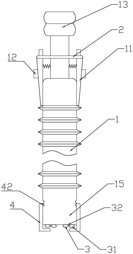
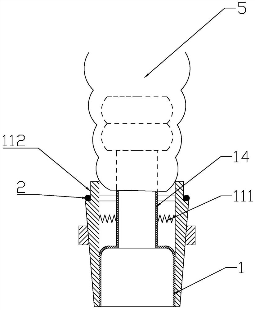
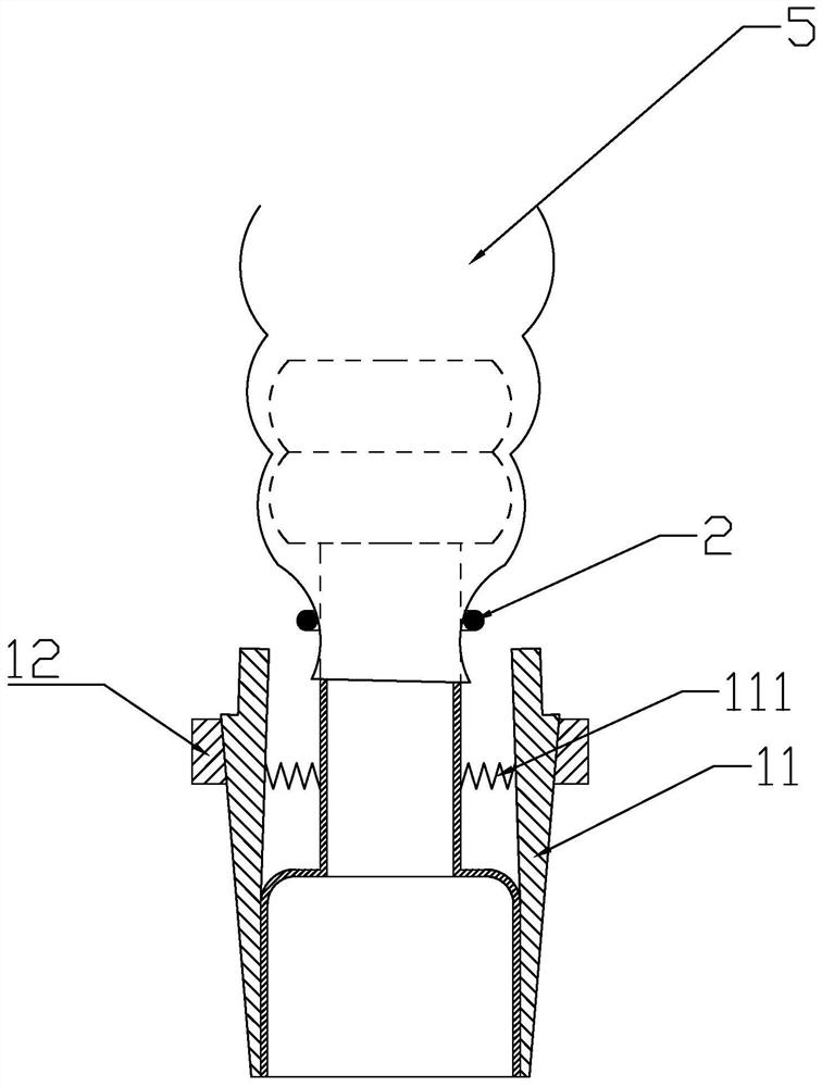
[0028] like figure 1 , figure 2 As shown, an intraoperative enteroscope intestinal wall fixation device includes a telescopic extension tube 1 made of plastic, and one end of the extension tube 1 is provided with a first joint for inserting into the colon 5, and the first joint includes a joint away from the extension tube 1. The outer section 13 of the outer section 13 and the inner section 14 close to the extension tube 1, the diameter of the inner section 14 is smaller than the diameter of the outer section 13 and the extension tube 1, and the outer section 13 has a wavy shape adapted to the shape of the inner wall of the colon 5. The extension tube 1 is provided with several elastic claws 11 along the circumference, one end of the elastic claws 11 is fixedly connected with the extension tube 1, the other end of the elastic claws 11 extend...


PUM


Abstract

The invention discloses an intestinal wall fixing device of anenteroscope during surgery. The intestinal wall fixing device comprises an extension tube, wherein one end of the extension tube is provided with a first connector used for being connected with the intestine in an inserted mode; a plurality of elastic claws are arranged on the extension tube along the circumferential direction; one end of the elastic claw is fixedly connected with the extension tube, and the other end of the elastic claw extend towards the direction of the first connector; the elastic claws are externally covered with an elastic ring; the other end of the extension tube is provided with a second connector used for being connected with the enteroscope in the inserted mode; and the second connector is provided with a sealing structure. By means of an elastic fixing device, knotting and fixing in the surgery are omitted, and surgery time is saved; by arrangement of the extension tube, the intestinal wall fixing device can be placed out of an operation field, more intestinal canals are prevented from being dissociated, surgery wounds are reduced, and the surgery time is shortened; by arrangement of a radial incision and an air bag, air leakage and intestinal content overflow are avoided, abdominal cavity and operation field pollution is avoided, and postoperative infection is avoided; after the endoscope enters during the surgery, suction, decompression and flushing can be carried out, the conditions of obstructed near-end intestinal canals are observed, and lesion omission is avoided; and the intestinal wall fixing device has a simple structure and is convenient in operation.

Description

technical field [0001] The invention relates to the technical field of medical devices, in particular to an intestinal wall fixation device for an intraoperative colonoscope. Background technique [0002] Intestinal obstruction is a common complication of colorectal cancer, and how to judge the lesion of the intestinal segment above the obstruction has always been a clinical problem. Intraoperative colonoscopy can directly understand the intestinal conditions above the obstruction site, and has clear guiding significance for surgical methods and surgical scope, especially for the discovery and surgical treatment of synchronous multiple colorectal cancer. At present, intraoperative colonoscopy routinely opens a hole in the proximal intestinal tract after tumor obstruction or cuts the intestinal tract to inspect the proximal intestinal tract. It is necessary to raise the proximal intestinal segment and place an incision outside the abdominal cavity, which will inevitably free ...

Claims


Application Information

Patent Timeline
Patent Type & Authority Applications(China)
IPC IPC(8): A61B1/31A61M1/00A61M3/02
CPCA61B1/00147A61B1/00082A61B1/31A61M3/02
Inventor 姚敏泉姜宇朋易兵鸿范金型
Owner 桐乡市第一人民医院
Learn More