
Brain tumor image segmentation method based on HDC-Net0 network

A brain tumor and image technology, applied in the field of brain tumor image segmentation, can solve the problems of insufficient segmentation accuracy of brain tumor images, limited computing overhead, GPU memory consumption, and low computing efficiency.

Inactive Publication Date: 2021-12-03
HENAN UNIVERSITY OF TECHNOLOGY
View PDF0 Cites 0 Cited by
  • Summary
  • Abstract
  • Description
  • Claims
  • Application Information

AI Technical Summary

Problems solved by technology

[0004] The content of the present invention is to solve the insufficient segmentation accuracy of brain tumor images and provide an automatic and accurate method for segmenting brain tumor images
[0006] (1) Aiming at the problems that the current 3D convolutional network has large computing memory and low computing efficiency, a lightweight pseudo-3D hierarchical decoupling network (HDC-Net 0 ) model, which completes the 3D segmentation task of brain tumors based on 2D convolution, and achieves excellent segmentation accuracy, which can effectively solve the problem of limited computing overhead and GPU memory consumption when performing brain tumor segmentation tasks based on 3D networks

Method used


Image

  • Brain tumor image segmentation method based on HDC-Net0 network
  • Brain tumor image segmentation method based on HDC-Net0 network

Examples


Embodiment Construction

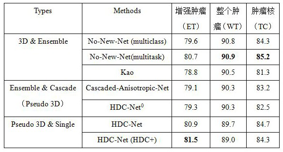
[0014] In order to verify the brain tumor image segmentation performance of the present invention, we selected the BraTS 2019 dataset for training and testing.

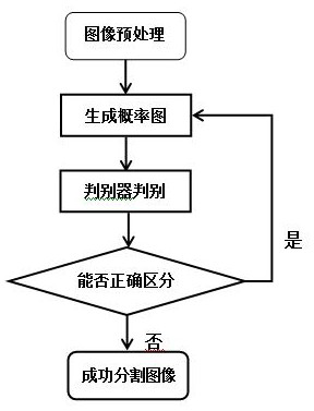
[0015] Step 1: Preprocessing the brain tumor image data, using Spyder software, using image rotation, translation transformation for image enhancement, and contrast enhancement for image normalization.

[0016] Step 2: Train HDC-Net in Spyder software 0 Network, during the training process, the patch size of the model input is 128×128×128, the Batch is 10, and the number of training is 800 epoch. The multi-category soft Dice function is used as the loss function of the model. There are two methods of data enhancement: random rotation and random brightness transformation. Before the image enters the network, it also uses the preprocessing method of 0 mean and 1 variance to adjust the gray scale range of the MR image.

[0017] Step 3: Use the test set of the BraTS 2019 dataset for HDC-Net 0 network for testing. It ...


PUM


Abstract

As is well known, malignant tumors are globally diseases which are difficult to treat and become one of the main diseases harming human life at present. Early accurate diagnosis, positioning, qualification, classification and segmentation of tumors are the key of follow-up treatment. The MRI brain tumor image segmentation means that a whole tumor area, a tumor core area and a tumor enhancement area are segmented from normal brain tissue. A traditional segmentation algorithm mainly challenges large gray similarity between brain tissues in an MRI image and differences between different cases. According to the multi-modal MRI brain tumor image segmentation, feature information of different modals in the MRI image can be fully utilized, the segmentation effectiveness is improved, and the multi-modal MRI brain tumor image segmentation is a research hotspot of brain tumor image processing in recent years.

Description

technical field [0001] The invention belongs to the technical field of medical image processing, and in particular relates to a segmentation method for brain tumor images. Background technique [0002] Brain tumor is one of the most common and extremely harmful diseases to the human body, with high morbidity and mortality. It has a strong social hazard. According to the survey, the number of brain tumor patients in my country is increasing year by year, and the situation is very serious. The magnetic resonance image analysis of brain tumors is an important basis for doctors to diagnose and treat brain tumors, evaluate surgical procedures, and track the condition. However, brain tumors have various shapes and complex structures, and manual segmentation is very time-consuming and labor-intensive, and it is easy to misjudgment during the segmentation process. Therefore, combining computer-aided diagnosis with computer vision technology, a fully automatic and precise segmentat...

Claims


Application Information

Patent Timeline
Patent Type & Authority Applications(China)
IPC IPC(8): G06T7/00G06T7/10G06T5/00G06T3/40G06T3/60G06N3/04G06N3/08
CPCG06T7/0012G06T7/10G06T3/4038G06T3/60G06N3/08G06T2207/20081G06T2207/20084G06T2207/20132G06T2207/30096G06N3/045G06T5/90
Inventor 李冰洁赵祥杨铁军张鑫
Owner HENAN UNIVERSITY OF TECHNOLOGY
Learn More