Chest CT image processing method and device

A CT image and processing method technology, applied in the computer field, can solve the problems of insufficiently precise lung disease information and affect the segmentation results, and achieve the effect of accurate extraction and precise diagnosis of information

Pending Publication Date: 2021-12-10
NORTHEASTERN UNIV
View PDF0 Cites 3 Cited by
  • Summary
  • Abstract
  • Description
  • Claims
  • Application Information

AI Technical Summary

Problems solved by technology

[0006] In view of the above-mentioned shortcomings and deficiencies of the prior art, the present invention provides a chest CT image processing method and device, which solves the problem that t...

Method used


Image

  • Chest CT image processing method and device
  • Chest CT image processing method and device
  • Chest CT image processing method and device

Examples


Embodiment Construction

[0143] In order to better explain the present invention and facilitate understanding, the present invention will be described in detail below through specific embodiments in conjunction with the accompanying drawings.

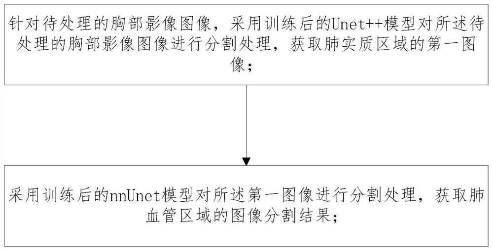
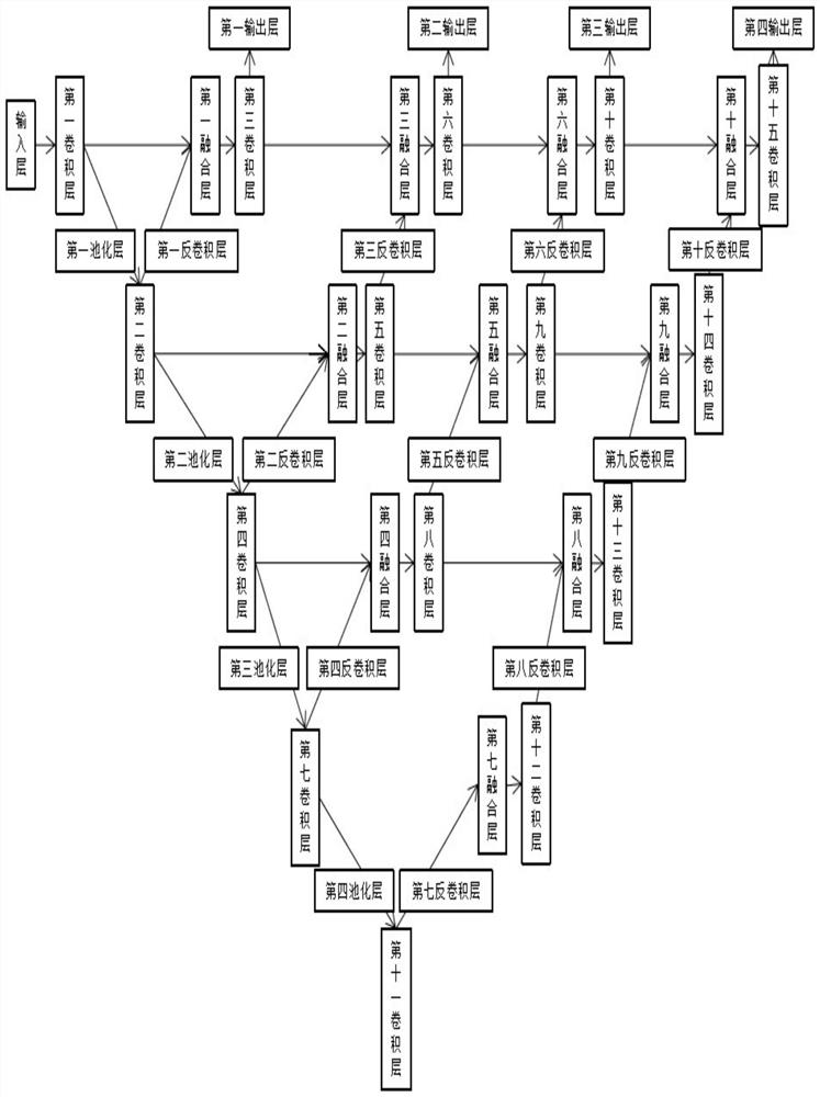
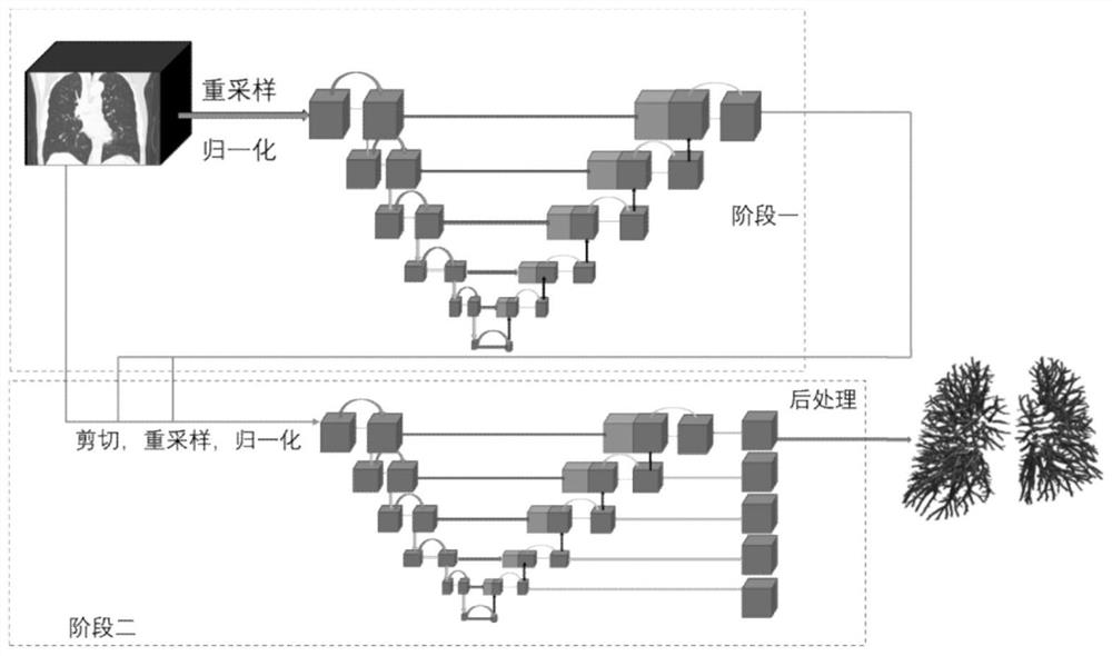
[0144] A chest CT image processing method and device proposed by the present invention, see figure 2 As shown, in the overall process, the lung parenchyma is extracted from the chest image to be processed first, and then the pulmonary blood vessels in the lung parenchyma image are extracted after the lung parenchyma in the image is accurately extracted, reducing the external Tissue interference increases the accuracy of extraction. In the extraction of lung parenchyma, a lung parenchyma segmentation algorithm based on the Unet++ network was implemented, and the network was trained to effectively reduce the over-segmentation rate. Afterwards, the pulmonary vessel segmentation algorithm based on nnUnet was used to extract the pulmonary vessels in the lung paren...


PUM


Abstract

The invention relates to a chest CT image processing method and device. The method comprises the steps: S01, carrying out segmentation processing of a to-be-processed chest image through a trained Unet++ model, and obtaining a first image of a pulmonary parenchyma region; and S02, performing segmentation processing on the first image by using a trained nnUnet model to obtain an image segmentation result of a pulmonary vessel region. Compared with the prior art, the method has the advantages that a full-automatic method for accurately and objectively separating pulmonary blood vessels based on conventional CT image analysis can be provided, the pulmonary blood vessels are accurately extracted, and finer diagnosis information is provided for doctors when doctors diagnose lung diseases.

Description

technical field [0001] The present invention relates to the field of computer technology, in particular to a chest CT image processing method and device. Background technique [0002] With the rapid growth of the world population and the arrival of aging, the form of cancer around the world is becoming more and more serious, and the incidence and mortality of cancer are increasing rapidly, among which the situation of lung cancer is particularly serious. In the process of treating lung cancer, doctors need to make diagnosis and treatment judgments on the patient's CT images first, but reading CT images requires strong professionalism, which is very complicated and difficult for non-professional doctors to read, so the pressure on doctors to read images will increase, resulting in doctors not getting more detailed information, and then unable to formulate more effective surgical plans and improve the survival rate of patients. [0003] The existing extraction of lung parench...

Claims


Application Information

Patent Timeline
IPC IPC(8): G06T7/00G06T7/11G06T5/50G06N3/04G06N3/08
CPCG06T7/0012G06T7/11G06T5/50G06N3/08G06T2207/10081G06T2207/20021G06T2207/20081G06T2207/20084G06T2207/20221G06T2207/30061G06T2207/30101G06N3/045
Inventor 徐士惠张子明邵慰周庆华王龙光魏泽旭韦祖懿焦树斌覃文军
Owner NORTHEASTERN UNIV
Why Eureka
Try Eureka