Blood vessel segmentation method and device, computer equipment and storage medium

A computer program and blood vessel technology, applied in the field of medical image processing, can solve the problems of under-segmentation and low segmentation accuracy, and achieve the effect of improving the accuracy

Pending Publication Date: 2022-02-08
SHANGHAI UNITED IMAGING INTELLIGENT MEDICAL TECH CO LTD +1
View PDF0 Cites 2 Cited by
  • Summary
  • Abstract
  • Description
  • Claims
  • Application Information

AI Technical Summary

Problems solved by technology

The existing blood vessel segmentation methods are affected by factors such as image quality and parameter selection, and there are problems of under-segmentation or over-segmentation, resulting in low segmentation accuracy

Method used


Image

  • Blood vessel segmentation method and device, computer equipment and storage medium
  • Blood vessel segmentation method and device, computer equipment and storage medium
  • Blood vessel segmentation method and device, computer equipment and storage medium

Examples


Embodiment Construction

[0070] In order to make the purpose, technical solution and advantages of the present application clearer, the present application will be further described in detail below in conjunction with the accompanying drawings and embodiments. It should be understood that the specific embodiments described here are only used to explain the present application, and are not intended to limit the present application.

[0071] The blood vessel segmentation method provided in this application can be applied to a terminal, and the terminal can be, but not limited to, a blood vessel imaging device, a personal computer, and the like.

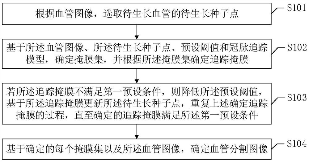
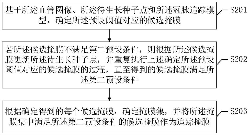
[0072] In one embodiment, such as figure 1 As shown, a blood vessel segmentation method is provided, and this embodiment is illustrated by applying the method to a terminal as an example. The method includes the following steps:

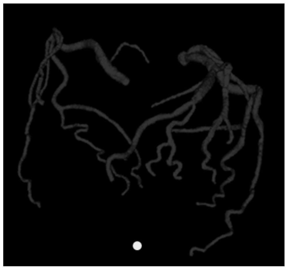
[0073] S101. Select a seed point of a blood vessel to be grown to be grown according to the blood vessel image.

[0074] Wherein, ...


PUM


Abstract

The invention relates to a blood vessel segmentation method and device, computer equipment and a storage medium. The method comprises the following steps: selecting a to-be-grown seed point of a to-be-grown blood vessel according to a blood vessel image; based on the blood vessel image, the to-be-grown seed point, a preset threshold value and a coronary artery tracking model, determining a mask set, and determining a tracking mask according to the mask set; if the tracking mask does not meet a first preset condition, reducing the preset threshold, updating the seed point to be grown based on the tracking mask, and repeating the process of determining the tracking mask until the determined tracking mask meets the first preset condition; and determining a blood vessel segmentation image based on each determined mask set and the blood vessel image. According to the invention, under-segmentation or over-segmentation caused by using a unified threshold value can be avoided, and the accuracy of blood vessel segmentation is improved.

Description

technical field [0001] The present application relates to the technical field of medical image processing, in particular to a blood vessel segmentation method, device, computer equipment and storage medium. Background technique [0002] The segmentation and extraction of blood vessels is an important prerequisite for the quantitative description of diseases and three-dimensional reconstruction of blood vessels, and it is also an important means to assist doctors in clinical diagnosis and treatment. Region growing methods are more common in vessel segmentation. [0003] The region growing method first sets a seed point, continuously traverses the neighborhood of the seed point, puts the points similar to the seed point into the same set, and then finds the similar points in the neighborhood of the midpoint of the set, and continuously expands the area until it is completed segmentation. The currently commonly used image segmentation methods based on seed points are generall...

Claims


Application Information

Patent Timeline
Patent Type & Authority Applications(China)
IPC IPC(8): G06T7/11G06T7/246G16H30/20
CPCG06T7/11G06T7/246G16H30/20G06T2207/30101
Inventor 杨帆董昢吴迪嘉李嘉豪雷丙震
Owner SHANGHAI UNITED IMAGING INTELLIGENT MEDICAL TECH CO LTD
Why Eureka
Try Eureka