Anatomy method for quickly exposing medical rat canalis spinalis
A medical and spinal canal technology, applied in the field of rat anatomy, can solve the problems of inability to quickly and successfully expose the spinal cord operation method, complicated and unsuitable for promotion, etc., and achieve the effect of suitable promotion, convenience and strong practicability
- Summary
- Abstract
- Description
- Claims
- Application Information
AI Technical Summary
Problems solved by technology
Method used
Image
Examples
Embodiment 1
[0022] Embodiment 1, the dissection method of the rapid exposure medical rat spinal canal comprises:
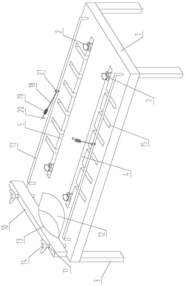
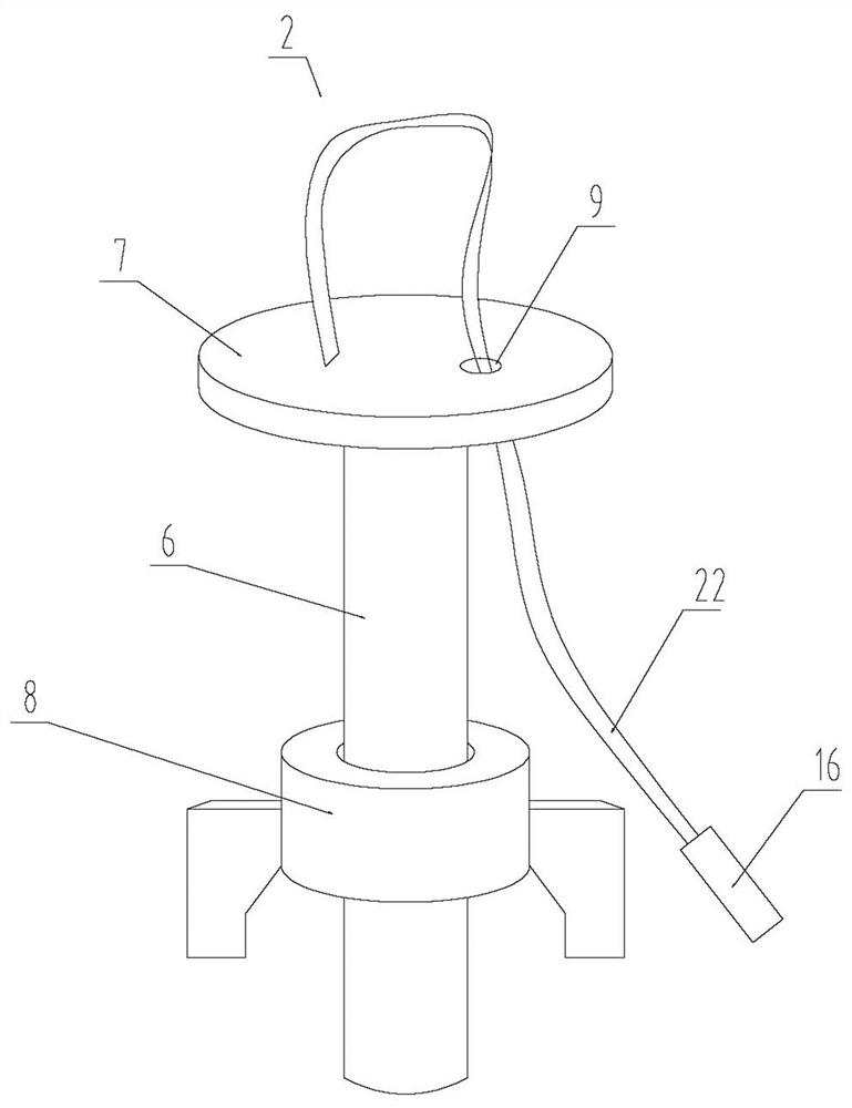
[0023] (1) Fixing the rats: select the dead rats, and fix the head and limbs of the rats on the experimental table through the fixing device to keep them in a prone position to ensure that the spines of the rats are in a straight state;
[0024] (2) Skin preparation in the surgical field: According to the needs of research and observation, select the target vertebrae of the corresponding segment. Taking the opening of the thoracic vertebral 12 spinal canal as an example, use scissors to cut off the rat hair around the back corresponding to the thoracic vertebral 12 spinal canal to reveal the rat. skin;
[0025] (3) Stripping tissue: Cut the back skin and subcutaneous soft tissue of the rat along the midline of the 12 spinous processes of the rat thoracic vertebrae with a sharp surgical knife to a depth of up to the spinous process, and then use a sharp surgical knife to strip...
Embodiment 2
[0027] Embodiment 2, the dissection method of the rapid exposure medical rat spinal canal includes:
[0028] (1) Fixing the rats: select the dead rats, and fix the head and limbs of the rats on the experimental table through the fixing device to keep them in a prone position to ensure that the spines of the rats are in a straight state;
[0029] (2) Skin preparation in the surgical field: According to the needs of research and observation, select the target vertebrae of the corresponding segment. Taking the opening of the thoracic vertebra 11 spinal canal as an example, use scissors to cut off the rat hair around the back corresponding to the thoracic vertebra 11 spinal canal to reveal the rat skin;
[0030] (3) Strip tissue: use a sharp surgical knife to incise the back skin and subcutaneous soft tissue of rats along the midline of the 11 spinous processes of the rat thoracic vertebrae to a depth of up to the spinous process, and then use a sharp surgical knife to peel off th...
Embodiment 3
[0032] Embodiment three, the dissection method of the rapid exposure medical rat spinal canal includes:
[0033] (1) Fixing the rats: select the dead rats, and fix the head and limbs of the rats on the experimental table through the fixing device to keep them in a prone position to ensure that the spines of the rats are in a straight state;
[0034] (2) Skin preparation in the surgical field: According to the needs of research and observation, select the target vertebrae of the corresponding segment. Taking the opening of the thoracic vertebral 12 spinal canal as an example, use scissors to cut off the rat hair around the back corresponding to the thoracic vertebral 12 spinal canal to reveal the rat. skin;
[0035] (3) Stripping tissue: Cut the back skin and subcutaneous soft tissue of the rat along the midline of the 12 spinous processes of the rat thoracic vertebrae with a sharp surgical knife to a depth of up to the spinous process, and then use a sharp surgical knife to st...
PUM
 Abstract
Description
Claims
Application Information
 - R&D Engineer
- R&D Manager
- IP Professional
- Industry Leading Data Capabilities
- Powerful AI technology
- Patent DNA Extraction
Browse by: Latest US Patents, China's latest patents, Technical Efficacy Thesaurus, Application Domain, Technology Topic, Popular Technical Reports.
© 2024 PatSnap. All rights reserved.Legal|Privacy policy|Modern Slavery Act Transparency Statement|Sitemap|About US| Contact US: help@patsnap.com


