
CT scanning method and device, electronic device and storage medium

A technology of CT scanning and CT value, which is applied in the field of medical imaging, can solve the problems of inaccuracy and other problems, and achieve the effect of avoiding dose increase, and the purpose and advantages are concise and easy to understand

Pending Publication Date: 2022-03-01
SHANGHAI UNITED IMAGING HEALTHCARE +1
View PDF0 Cites 5 Cited by
  • Summary
  • Abstract
  • Description
  • Claims
  • Application Information

AI Technical Summary

Problems solved by technology

[0006] In this embodiment, a CT scanning method, device, electronic device and storage medium are provided to solve the problem that in the multi-phase or multi-part enhanced scan in the related art, the traditional threshold tracking technology can only measure the region of interest. The CT value is used to determine the scan time, and the regions of interest in other parts or phases can only be judged by experience, which is not accurate enough

Method used


Image

  • CT scanning method and device, electronic device and storage medium
  • CT scanning method and device, electronic device and storage medium
  • CT scanning method and device, electronic device and storage medium

Examples


Embodiment Construction

[0030] In order to understand the purpose, technical solution and advantages of the present application more clearly, the present application is described and illustrated below in conjunction with the accompanying drawings and embodiments.

[0031] Unless otherwise defined, the technical terms or scientific terms involved in the application shall have the general meanings understood by those skilled in the technical field to which the application belongs. In this application, words like "a", "an", "an", "the", "these" and the like do not denote quantitative limitations, and they may be singular or plural. The terms "comprising", "comprising", "having" and any variants thereof referred to in this application are intended to cover non-exclusive inclusion; for example, processes, methods and The system, product or device is not limited to the steps or modules (units) listed, but may include steps or modules (units) not listed, or may include other steps or modules inherent to the...


PUM


Abstract

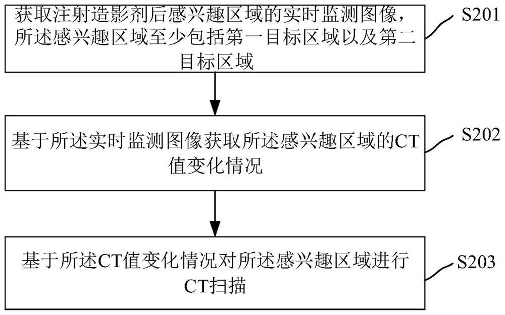
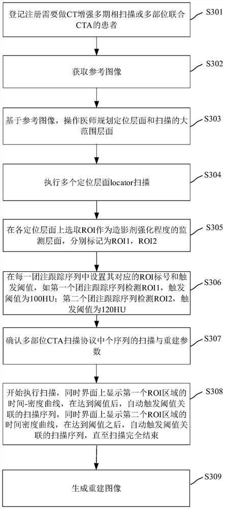
The invention relates to a CT scanning method, and the method comprises the steps: obtaining a real-time monitoring image of a region of interest after a contrast agent is injected, and the region of interest at least comprises a first target region and a second target region; acquiring a CT value change condition of the region of interest based on the real-time monitoring image; and performing CT scanning on the region of interest based on the CT value change condition. According to the method and the device, the problems that in multi-phase or multi-part enhanced scanning in related technologies, a traditional threshold tracking technology can only measure the CT value of one part or phase to determine the scanning time, and other parts or phases can only be judged depending on experience and are not accurate enough are solved. According to the method, the scanning triggering time point with the best scanning effect of each part can be accurately determined in multi-part or multi-phase enhanced scanning, the optimal image data can be obtained, and the risk of dosage increase caused by repeated scanning due to inaccurate scanning triggering time points can be avoided.

Description

technical field [0001] The present application relates to the technical field of medical imaging, in particular to a CT scanning method, device, electronic device and storage medium. Background technique [0002] Computer tomography (Computed Tomography, CT) uses precisely collimated X-ray beams, γ-rays, ultrasound, etc., together with highly sensitive detectors, to conduct cross-sectional scans one after another around a certain part of the human body. Fast, clear images, etc., can be used for the examination of various diseases; according to the different rays used, it can be divided into: X-ray CT (X-CT) and γ-ray CT (γ-CT). [0003] CTA is the abbreviation of CT angiography (CT angiography). Spiral CT scanning is performed after intravenous injection of contrast agent. During 3D reconstruction, structures that do not need to be displayed, such as skin, muscle, and bone, are removed, and only the 3D vascular structure and visceral structure are displayed. CTA has been Wi...

Claims


Application Information

Patent Timeline
IPC IPC(8): A61B6/00A61B6/03
CPCA61B6/481A61B6/5211A61B6/032
Inventor 李真林祝雨雷
Owner SHANGHAI UNITED IMAGING HEALTHCARE
Learn More