Heart image acquisition method and device, magnetic resonance equipment and storage medium

An image acquisition and magnetic resonance technology, which is applied in magnetic resonance measurement, measurement using nuclear magnetic resonance imaging system, magnetic variable measurement, etc. Effect

Pending Publication Date: 2022-03-01
UNITED IMAGING RES INST OF INNOVATIVE MEDICAL EQUIP
View PDF0 Cites 0 Cited by
  • Summary
  • Abstract
  • Description
  • Claims
  • Application Information

AI Technical Summary

Problems solved by technology

[0004] However, the current ECG-triggered magnetic resonance heart scanning technology has the problem of not being able to obtain systolic images

Method used


Image

  • Heart image acquisition method and device, magnetic resonance equipment and storage medium
  • Heart image acquisition method and device, magnetic resonance equipment and storage medium
  • Heart image acquisition method and device, magnetic resonance equipment and storage medium

Examples


Embodiment Construction

[0044] In order to make the purpose, technical solution and advantages of the present application clearer, the present application will be further described in detail below in conjunction with the accompanying drawings and embodiments. It should be understood that the specific embodiments described here are only used to explain the present application, and are not intended to limit the present application.

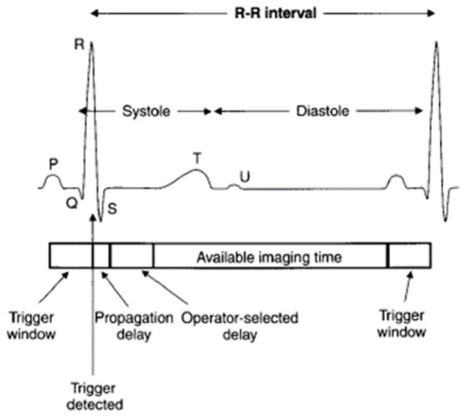
[0045] In the prior art, when performing magnetic resonance imaging on the heart, electrocardiographic triggering is commonly used, that is, by attaching electrodes on the surface of the target object to measure electrocardiographic signals during the scanning process of the MRI equipment. Specifically, when the ECG signal is displayed in the systolic phase, the MRI scan is stopped, and when the ECG signal is displayed in the diastolic phase, the MRI scan is started. figure 1 Schematic diagram of available imaging times for conventional ECG triggering. Since the prior art...


PUM


Abstract

The invention relates to a heart image acquisition method and device, magnetic resonance equipment and a storage medium. The method comprises the following steps: acquiring a preset excitation space of a target scanning layer of a heart area, determining a target radio frequency pulse according to parameters of the preset excitation space and a preset gradient, exciting the preset excitation space by using the target radio frequency pulse, sampling the excited excitation space, filling a two-dimensional k space with sampling points, and obtaining a target scanning layer of the heart area; according to the method, the two-dimensional k space is subjected to image reconstruction to obtain the magnetic resonance heart image, the size of the original view space is reduced, it is ensured that the image in the systolic period can be scanned, and the two-dimensional k space is selected, so that motion artifacts generated during systole when a traditional three-dimensional k space is excited for scanning can be avoided.

Description

technical field [0001] The present application relates to the field of magnetic resonance technology, in particular to a cardiac image acquisition method, device, magnetic resonance equipment and storage medium. Background technique [0002] Magnetic resonance imaging (MRI) systems are widely used in medical diagnosis. For example, Cardiac Magnetic Resonance Imaging (CMR) technology is widely used in routine examination of a patient's heart. [0003] At present, when using MRI equipment to scan the heart, ECG triggering is generally used for scanning, that is, by sticking electrodes on the body surface, when the MRI equipment detects that the ECG signal is displayed in the systole during the scanning process, the MRI equipment stops. Scanning, when the ECG signal shows that the heart is diastolic, the MRI machine starts scanning. [0004] However, the current electrocardiographically triggered magnetic resonance heart scanning technology has the problem of not being able t...

Claims


Application Information

Patent Timeline
Patent Type & Authority Applications(China)
IPC IPC(8): G01R33/48
CPCG01R33/48
Inventor 全希佳
Owner UNITED IMAGING RES INST OF INNOVATIVE MEDICAL EQUIP
Why Eureka
Try Eureka