
Ultrasonic endoscope

A technology of ultrasound and endoscopy, applied in the field of ultrasound endoscopy, can solve problems such as unevenness and image quality degradation, and achieve the effect of reducing diameter and suppressing image quality degradation

Pending Publication Date: 2022-03-08
FUJIFILM CORP
View PDF1 Cites 0 Cited by
  • Summary
  • Abstract
  • Description
  • Claims
  • Application Information

AI Technical Summary

Problems solved by technology

However, when a plurality of signal lines included in a non-coaxial cable are connected to an ultrasonic vibrator, image quality degradation (for example, unevenness) may occur in an ultrasonic image due to the length of the signal line.

Method used


Image

  • Ultrasonic endoscope
  • Ultrasonic endoscope
  • Ultrasonic endoscope

Examples


Embodiment Construction

[0029] Hereinafter, preferred embodiments of the ultrasonic endoscope according to the present invention will be described with reference to the drawings.

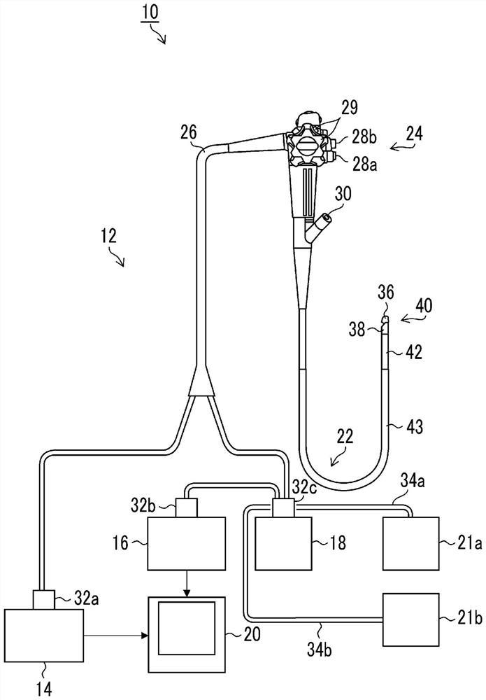
[0030] figure 1 It is a schematic configuration diagram showing an example of an ultrasonic examination system 10 using the ultrasonic endoscope 12 according to the embodiment.

[0031] like figure 1 As shown, the ultrasonic examination system 10 includes: an ultrasonic endoscope 12; an ultrasonic processor device 14 for generating an ultrasonic image; an endoscope processor device 16 for generating an endoscopic image; a light source device 18 for providing an ultrasonic endoscope 12 supplies illumination light to illuminate the body cavity; and a display 20 displays ultrasonic images and endoscopic images. Furthermore, the ultrasonic examination system 10 includes: a water supply tank 21a for storing washing water and the like; and a suction pump 21b for suctioning the aspirated matter in the body cavity.

[0032] The...


PUM


Abstract

An ultrasonic endoscope is provided with: an insertion section including a tip section having an ultrasonic transducer array in which a plurality of ultrasonic transducers are arranged; a cable inserted through the insertion part; a substrate that electrically connects the plurality of ultrasonic transducers and a cable and is disposed at the tip portion, the cable having: a non-coaxial cable including a first cable bundle comprising a plurality of signal lines and a plurality of ground lines, and a first shield layer covering the first cable bundle; and a sheath covering a second cable bundle comprising a plurality of non-coaxial cables, the substrate having a plurality of electrode pads connected to the plurality of ultrasonic transducers, the first cable bundles being led out from the cables, the signal lines of the first cable bundles being led out and electrically connected to the electrode pads of the substrate, and the signal lines of the second cable bundles being led out and electrically connected to the electrode pads of the substrate. The plurality of ultrasonic vibrators are configured from a plurality of drive units that are simultaneously driven, the plurality of signal lines of each first cable bundle are connected to the plurality of electrode pads of each drive unit to constitute a signal line group, and the signal line group includes at least two types of signal lines having different lengths from the tip of the first cable bundle.

Description

technical field [0001] The invention relates to an ultrasonic endoscope. Background technique [0002] In recent years, ultrasonic endoscopes that irradiate the internal body of a subject with ultrasonic waves, receive the reflected waves and visualize them to observe the state of the internal body have been used in medical fields. [0003] Such an ultrasonic endoscope, for example, as disclosed in Patent Document 1, includes: a distal end portion provided with a piezoelectric element constituting an ultrasonic vibrator; a bending portion and a flexible portion connected to the proximal end of the distal end portion; The axial cable is inserted and penetrated through the bending portion and the flexible portion; and the wiring board is electrically connected to the piezoelectric element and the coaxial cable. [0004] Patent Document 1: Japanese Patent Laid-Open No. 2019-054962 [0005] Among them, in the case of a coaxial cable, since a shielding layer and a sheath cover ...

Claims


Application Information

Patent Timeline
Patent Type & Authority Applications(China)
IPC IPC(8): A61B8/12A61B8/00
CPCA61B8/12A61B8/42A61B8/56A61B8/4483A61B8/4494A61B8/445B06B1/06B06B1/0207A61B2562/182
Inventor 岩屋达广森本康彦
Owner FUJIFILM CORP
Learn More