Sleep state detection method, device and system based on electroencephalogram acquisition head ring
A sleep state and detection method technology, applied in the field of sleep detection, can solve problems such as poor test results, and achieve the effects of correct classification, accurate collection, and accurate identification
- Summary
- Abstract
- Description
- Claims
- Application Information
AI Technical Summary
Problems solved by technology
Method used
Image

Examples
Embodiment 1
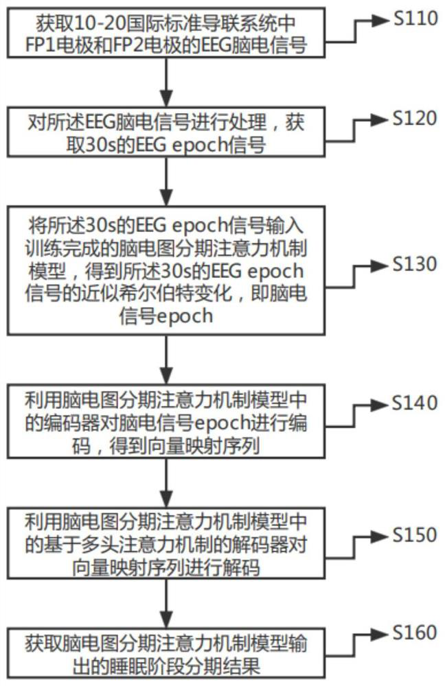
[0079] figure 1 This is a schematic flowchart of the sleep state detection method based on the EEG acquisition headband provided in the first embodiment of the present invention. This embodiment can be applied to the sleep state detection using the portable EEG acquisition headband. The sleep state detection device is implemented, which specifically includes the following steps:
[0080] S110. Obtain the EEG signals of the FP1 electrode and the FP2 electrode in the 10-20 international standard lead system;
[0081] Exemplarily, a portable EEG signal acquisition headband can be used, and electrode pads can be placed with reference to the 10-20 system electrode placement method to obtain the EEG EEG of the user's FP1 and FP2 electrodes corresponding to the 10-20 system electrode placement method. Signal,
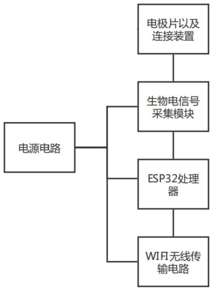
[0082] figure 2 For a schematic diagram of the structure of the EEG signal acquisition head ring in the sleep state detection method based on the EEG acquisition head ring...
Embodiment 2
[0115] Figure 7 This is a schematic flowchart of a sleep state detection method based on an EEG acquisition headband provided in Embodiment 2 of the present invention; this embodiment is optimized on the basis of the above-mentioned embodiment. In this embodiment, the EEG phased attention mechanism is used. The encoder in the model encodes the EEG signal epoch, and after obtaining the vector mapping sequence, the following steps are added: input the vector mapping sequence output by the global average pooling layer into the first DropOut layer to obtain the output of the encoder; The DropOut probability of the DropOut layer is 0.5.
[0116] Correspondingly, the sleep state detection method based on the EEG acquisition headband provided by this embodiment specifically includes:
[0117] S210. Obtain the EEG signals of the FP1 electrode and the FP2 electrode in the 10-20 international standard lead system;
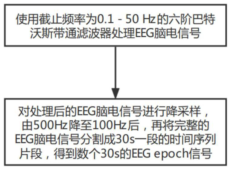
[0118] S220, processing the EEG signal to obtain an EEG epoch signal...
Embodiment 3
[0140] Figure 9 This is a schematic flowchart of a sleep state detection method based on an EEG acquisition headband provided in Embodiment 3 of the present invention; this embodiment is optimized on the basis of the above-mentioned embodiment. In this embodiment, the output of the encoder is encoded with the position After the PE is summed and normalized, and then input to the multi-head attention mechanism sub-module of the decoder, the following steps are added: Input the output of the multi-head attention mechanism sub-module to the third DropOut layer, and the DropOut probability of the third DropOut layer is 0.8 , the output of the multi-head attention mechanism sub-module is normalized through the processing of the third DropOut layer; among them, the number of heads in the multi-head attention mechanism sub-module is 8, the size of each head is 64 dimensions, and the attention mechanism is in each The heads are executed in parallel; the attention mechanism function is...
PUM
 Abstract
Description
Claims
Application Information
PatSnap Eureka turns technology decisions into work you can execute. Powered by our Innovation Knowledge Graph, it runs expert workflows across engineering, life sciences, materials and intellectual property. Get your review-ready output in minutes.


