
Pulmonary vessel segmentation method and device, storage medium and electronic equipment

A technology for blood vessels and segments to be segmented, applied in the field of image processing, can solve problems such as stitching traces at stitching places, achieve the effect of improving accuracy and optimizing segmentation effect

Pending Publication Date: 2022-06-07
INFERVISION MEDICAL TECH CO LTD
View PDF0 Cites 2 Cited by
  • Summary
  • Abstract
  • Description
  • Claims
  • Application Information

AI Technical Summary

Problems solved by technology

The above-mentioned segmentation method for lung CT images will lead to obvious stitching marks at the stitching places, especially at the stitching places related to the mediastinum, where the stitching marks are more obvious

Method used


Image

  • Pulmonary vessel segmentation method and device, storage medium and electronic equipment
  • Pulmonary vessel segmentation method and device, storage medium and electronic equipment
  • Pulmonary vessel segmentation method and device, storage medium and electronic equipment

Examples


Embodiment Construction

[0029] The technical solutions in the embodiments of the present application will be clearly and completely described below with reference to the accompanying drawings in the embodiments of the present application. Obviously, the described embodiments are only a part of the embodiments of the present application, but not all of the embodiments. Based on the embodiments in the present application, all other embodiments obtained by those of ordinary skill in the art without creative efforts shall fall within the protection scope of the present application.

[0030] CT (Computed Tomography), that is, electronic computed tomography, which uses precisely collimated X-ray beams, gamma rays, ultrasonic waves, etc., together with a highly sensitive detector, performs cross-sectional scans one by one around a certain part of the human body. It has the characteristics of fast scanning time and clear images, and can be used for the inspection of various diseases.

[0031] The lung consis...


PUM


Abstract

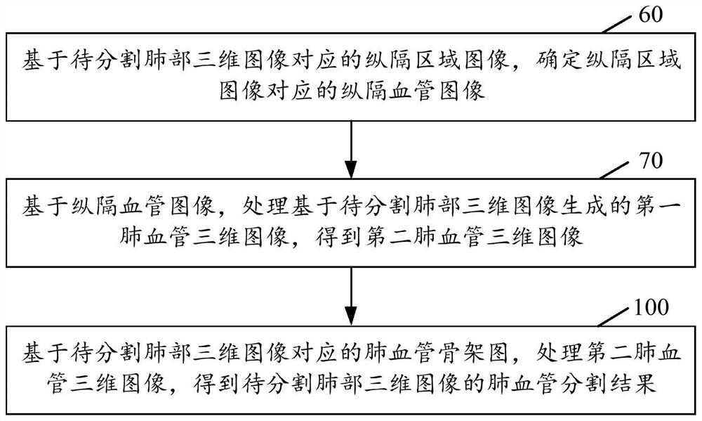
The invention provides a pulmonary vessel segmentation method and device, a storage medium and electronic equipment, and relates to the technical field of image processing. The lung blood vessel segmentation method comprises the steps of determining a mediastinal blood vessel image corresponding to a mediastinal region image based on the mediastinal region image corresponding to a to-be-segmented lung three-dimensional image; based on the mediastinal blood vessel image, processing a first lung blood vessel three-dimensional image generated based on the to-be-segmented lung three-dimensional image to obtain a second lung blood vessel three-dimensional image; and processing the second lung blood vessel three-dimensional image based on the lung blood vessel skeleton diagram corresponding to the lung three-dimensional image to be segmented to obtain a lung blood vessel segmentation result of the lung three-dimensional image to be segmented. According to the pulmonary vessel segmentation method, the segmentation result of the mediastinum is optimized, so that the final pulmonary vessel segmentation result is more accurate, and doctors can conveniently diagnose lung-related diseases of patients.

Description

technical field [0001] The present application relates to the technical field of image processing, and in particular, to a pulmonary blood vessel segmentation method, device, storage medium and electronic device. Background technique [0002] In order to better assist doctors in the diagnosis and treatment of lung-related diseases with the help of lung medical images, it is usually necessary to perform blood vessel segmentation on lung medical images to obtain pulmonary blood vessel segmentation results. [0003] However, due to the characteristics of dense and slender pulmonary blood vessels, the current segmentation methods for pulmonary blood vessels have the defect of poor segmentation effect. For example, for the segmentation method of Computed Tomography (CT) images of the lungs, it is usually necessary to slice the CT images of the lungs first to obtain multiple lung slices, and then use the deep learning model to segment each lung separately. The lung segments are s...

Claims


Application Information

Patent Timeline
IPC IPC(8): G06T7/00G06T7/11G06T7/136G06T7/187
CPCG06T7/0012G06T7/11G06T7/136G06T7/187G06T2207/10081G06T2207/30061G06T2207/30101
Inventor 郝增号刘恩佑张欢陈宽王少康
Owner INFERVISION MEDICAL TECH CO LTD
Learn More