
Cornea edge stem cell tissue engineering composite body and its preparation method

A corneal limbal stem cell and tissue engineering technology is applied in the field of human corneal limbal stem cell-amniotic membrane tissue engineering complex and its preparation to achieve the effect of promoting cell differentiation

Inactive Publication Date: 2005-03-09
天津医科大学眼科中心
View PDF0 Cites 17 Cited by
  • Summary
  • Abstract
  • Description
  • Claims
  • Application Information

AI Technical Summary

Problems solved by technology

However, traditional surgical methods such as palpebral suture, autologous conjunctival transplantation, oral mucosal transplantation, amniotic membrane transplantation, and corneal limbal stem cell transplantation all have many limitations. The cure of severe corneal limbal stem cell-deficient ocular surface diseases urgently requires new treatment techniques and therapeutic products

Method used


Image

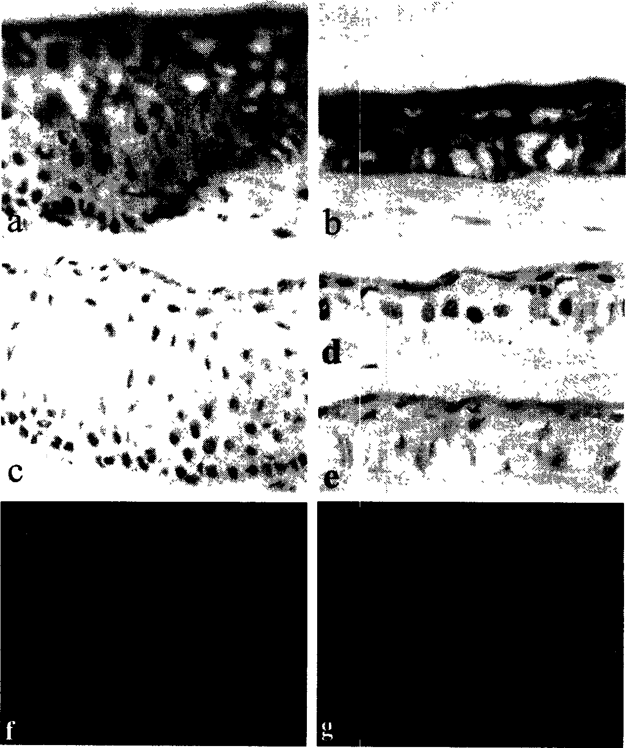
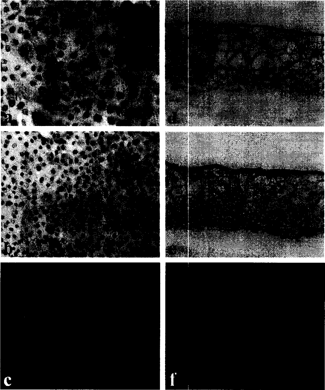
  • Cornea edge stem cell tissue engineering composite body and its preparation method
  • Cornea edge stem cell tissue engineering composite body and its preparation method
  • Cornea edge stem cell tissue engineering composite body and its preparation method

Examples


Embodiment Construction

[0025] Below, the present invention will be further described in conjunction with the examples, but the present invention is not limited to the following examples.

[0026] 1. Use the following methods to culture human limbal stem cells-amnion complex:

[0027] 1) Preparation of cultured film

[0028] The cesarean section fetal membranes of healthy pregnant women were taken, and the amniotic membrane and chorion were separated along the physiological gap, and the amniotic membrane epithelium was placed on the nitrocellulose membrane, and placed in a composition composed of 50% DMEM (Gibco, USA) and 50% glycerin stored at -80°C for more than three months. When in use, take out the frozen amnion from the refrigerator and put it in a 37°C water bath to thaw. Act with 0.1% EDTA for 30 minutes, then act with 0.1% trypsin for 30 seconds, and gently scrape off the amnion epithelial cells with a cell scraper. Lay the amniotic epithelium face up in the culture insert (Millipore, USA...


PUM


Abstract

A tissue-engineered complex of human limbus stem cell and amnion for treating the limbus stem cell-deficiency eye diseases is disclosed. It is perpared through removing epithelium from amnion, using it as carrier, using fibroblast 3T3 as nutritive layer, culturing by cell suspension method, and promoting differentiation of cells by air-lifting technique.

Description

(1) Technical field: [0001] The invention relates to a human corneal limbal stem cell-amnion tissue engineering complex capable of repairing limbal stem cell damage and used for ocular surface reconstruction and its preparation technology, in particular to a limbal stem cell tissue engineering complex and a preparation method thereof. (2) Technical background: [0002] In the treatment of ocular surface diseases in recent years, the most important breakthrough is the understanding of the function and localization of limbal stem cells. A large number of research results have shown that some cells in the basal layer of the limbus maintain undifferentiated characteristics and are "stem cells" of corneal epithelial cells. These cells have the following characteristics: (1) have proliferation potential, (2) can self-renew, (3) ) can produce a large number of terminally differentiated functional cells, and (4) can repair tissue damage. Ocular surface diseases characterized by lim...

Claims


Application Information

Patent Timeline
IPC IPC(8): C12N5/074
Inventor 孙慧敏张晓敏李筱荣
Owner 天津医科大学眼科中心
Learn More