

X-ray computerised tomograph capable of automatic eliminating black false image
A computer and black-hearted technology, which is applied in the field of medical image processing, can solve the problems of artifact image and real image size mismatch, affecting image quality, and different reconstruction field of view, etc.
- Summary
- Abstract
- Description
- Claims
- Application Information
AI Technical Summary
Problems solved by technology
Method used
Image

Examples
Embodiment 1
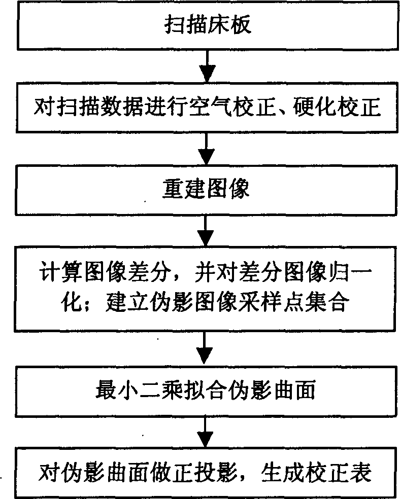
[0055] Implement the present invention on a double-layer CT machine, scan bed board data, bed height 445mm, body scanning protocol 120KV, 150mA, minimum scanning time 2 seconds, slice thickness 7mm, tomographic scanning mode.
[0056]After the system automatically performs air correction and hardening correction, the raw data Raw is obtained, and the raw data Raw is reconstructed by convolution and back-projection to obtain the bed board image CouchImg. Because the tube is at the zero position, the differential operation is performed along the image from top to bottom, and the CouchImg differential image is calculated to obtain CouchImg'.
[0057] CouchImg(i,j)=CouchImg(i+1,j)-CouchImg(i,j),
[0058] Wherein, i=0, .., IMGSIZE-1, j=0, . . . , IMGSIZE, i, j correspond to directions parallel to and perpendicular to the bed board, respectively.
[0059] Normalized difference image CouchImg normalized operation
[0060] CouchIm g ′ ...
Embodiment 2
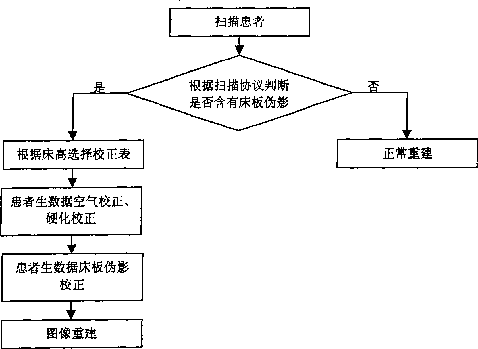
[0073] The invention was implemented on a single slice CT machine. Scan bed board data, bed height 400mm, body scan protocol 80KV, 35mA, minimum scan time 5 seconds, slice thickness 10mm, tomographic scan mode. After the system automatically performs air correction and hardening correction, the raw data Raw is obtained, and the algebraic reconstruction algorithm is used to obtain the bed board image CouchImg on the raw data. Because the tube is at the zero position, the difference calculation is performed along the image from top to bottom, and the CouchImg difference image is calculated to obtain CouchImg'.
[0074] CouchImg(i,j)=CouchImg(i+1,j)-CouchImg(i,j),
[0075] Wherein, i=0, .., IMGSIZE-1, j=0, . . . , IMGSIZE, i, j correspond to directions parallel to and perpendicular to the bed board, respectively.
[0076] Normalized difference image CouchImg normalized operation
[0077] CouchIm g ′ 1 ( ...
Embodiment 3
[0088] The invention is implemented on a multi-slice CT machine, the bed board data is scanned, the bed height is 550 mm, the body scanning protocol is 140 KV, 300 mA, the shortest scanning time is 0.8 seconds, the slice thickness is 1 mm, and the tomographic scanning mode is used. After the system automatically performs air correction and hardening correction, the raw data Raw data is obtained for Fourier reconstruction algorithm reconstruction, and the bed plate image is obtained. Because the tube is at the zero position, the differential operation is performed along the image from top to bottom, and the CouchImg differential image is calculated to obtain CouchImg'.
[0089] CouchImg(i,j)=CouchImg(i+1,j)-CouchImg(i,j),
[0090] Wherein, i=0, .., IMGSIZE-1, j=0, . . . , IMGSIZE, i, j correspond to directions parallel to and perpendicular to the bed board, respectively.
[0091] Normalized difference image CouchImg normalized operation
[0092] CouchIm ...
PUM
 Abstract
Description
Claims
Application Information