
X-ray diagnostic imaging system and x-ray diagnostic imaging method

An image diagnosis and X-ray technology, which is applied in the fields of radiological diagnostic equipment, diagnosis, medical science, etc., can solve the problems of increased invasiveness, increased patient burden, and increased X-ray exposure, and achieve the goal of suppressing invasiveness Effect

Inactive Publication Date: 2006-01-25
KK TOSHIBA +1
View PDF0 Cites 5 Cited by
  • Summary
  • Abstract
  • Description
  • Claims
  • Application Information

AI Technical Summary

Problems solved by technology

Since the contrast medium is invasive, the invasiveness of the patient increases by performing two contrast medium injections, and the burden on the patient increases
[0012] In addition, if the patient is injected with contrast medium twice and the imaging of the mask image is performed twice, it will take a lot of imaging time, so the amount of X-ray exposure to the patient will increase, and the work efficiency of the diagnosis and treatment will be reduced.

Method used


Image

  • X-ray diagnostic imaging system and x-ray diagnostic imaging method
  • X-ray diagnostic imaging system and x-ray diagnostic imaging method
  • X-ray diagnostic imaging system and x-ray diagnostic imaging method

Examples


Embodiment Construction

[0024] With reference to the drawings, the X-ray image diagnosis device and the diagnosis method related to the present invention

[0025] The embodiment will be described.

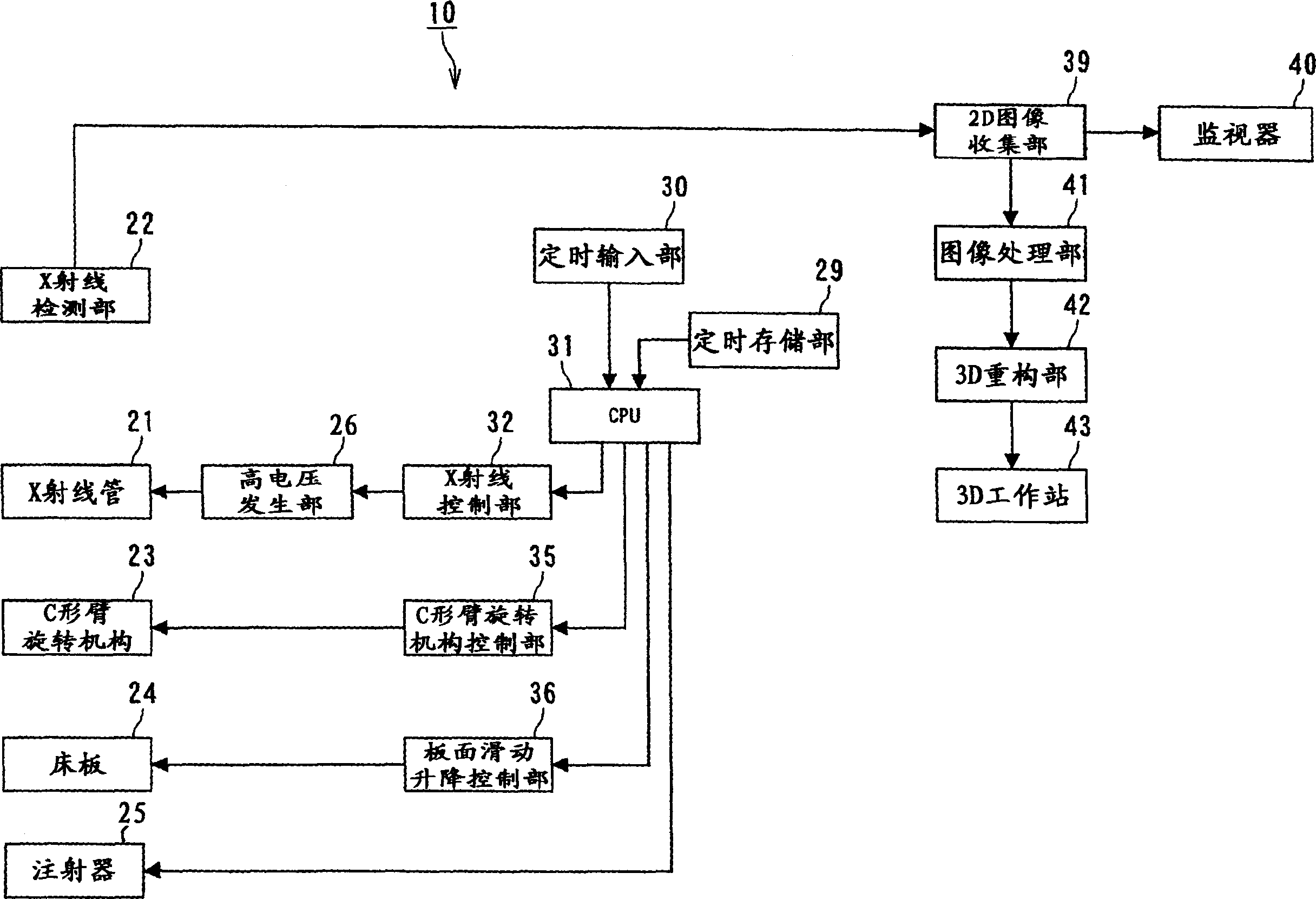
[0026] figure 1 It is a block diagram showing an embodiment of the X-ray image diagnosis apparatus related to the present invention.

[0027] figure 1 As an example of an X-ray image diagnosis apparatus, a 3D (three-dimensional) vasculature will be described. For image creation in 3D vasculature, for example, there is a DA (Digital Angiography) imaging mode in which general X-ray imaging is performed, which simply collects image data of X-ray images containing (the flow of) a contrast agent, and displays and stores it.

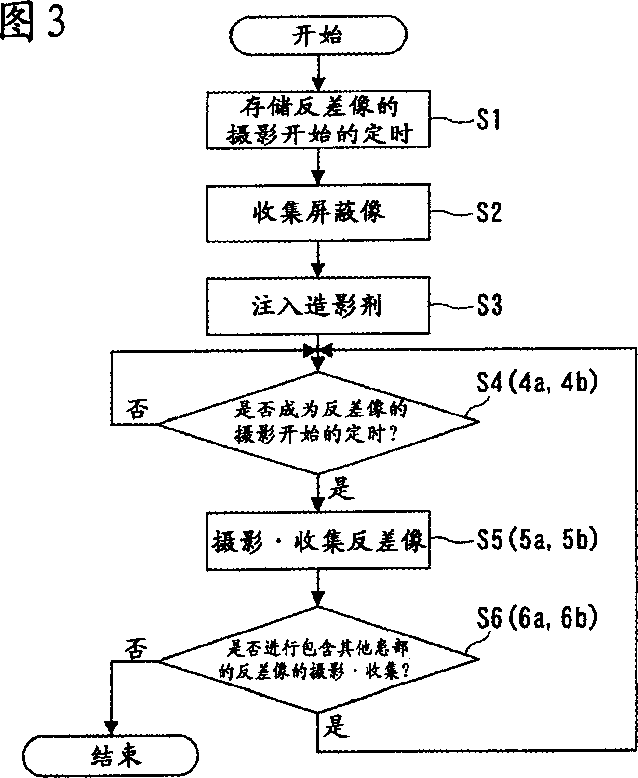
[0028] In addition, there is image data that subtracts the X-ray image of the image containing no contrast agent (shield image) and the X-ray image (contrast image or live image) of the image containing the contrast agent to generate a difference image. This is a DSA (Digital Subtraction An...


PUM


Abstract

The invention provides an X-ray image diagnostic device (10) used to photograph the necessary parts of patient P, collect all the image data of the shielded image and contrast image, and record and display the image data of the differential image of the shielded image and the contrast image. The device consists of a memory-timing part(39) and a CPU (31), wherein the memory-timing part(39) is used to record the timing of the start of the photograph of a first contrast image and a second contrast image; the CPU (31) is used to give instructions to carry out the photograph of the contrast image after the photograph of the contrast image to be stored at the timed memory part(39) starts. As the contrast medium is injected into the affected part, according to the instructions of the CPU (31), the photograph and the collection of the first contrast image is carried out, and then according to instructions of the CPU (31), the photograph and the collection of the second contrast image are carried out.

Description

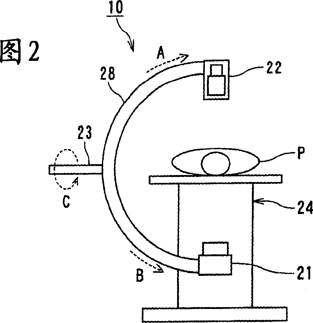
Technical field [0001] The present invention relates to an X-ray image diagnosis apparatus and a diagnosis method for imaging the internal structure of a patient by using a contrast agent. Background technique [0002] In order to photograph blood vessels whose contrast has been enhanced with a contrast agent, for example, X-ray tubes and detectors are provided at both ends of a substantially C-shaped support (hereinafter referred to as "C-arm") {II(Image An X-ray image diagnostic device composed of an Intensifier, a planar detector, etc., and an image processing unit is already known to the public. This X-ray image diagnosis apparatus is also generally called a vasculature device, and can be diagnosed, treated (diagnosed and treated) or X-ray diagnosed by a doctor who performs catheterization work on a patient. [0003] In diagnosis and treatment performed by doctors and other operators, fluoroscopy using X-ray image diagnosis equipment is used. In this fluoroscopy, the captured...

Claims


Application Information

Patent Timeline
IPC IPC(8): A61B6/00
Inventor 山田尚树
Owner KK TOSHIBA
Learn More