
Multiplicated imaging system

An imaging system and multiple technologies, applied in medical science, sensors, diagnostic recording/measurement, etc., can solve the problems of unequal deformation, time-consuming and laborious, spatial coordinate changes, etc., to shorten inspection time, improve simplicity, and reduce fear. sense of effect

Active Publication Date: 2006-04-19
SHENYANG NEUSOFT PAISITONG MEDICAL SYST
View PDF4 Cites 20 Cited by
  • Summary
  • Abstract
  • Description
  • Claims
  • Application Information

AI Technical Summary

Problems solved by technology

[0010] The second is that the multiple imaging systems have been registered to the same position on the axis, but due to the limited rigidity of the bed board and bed material, this makes them bear the weight of the diagnosed person in each imaging subsystem of the multiple imaging system. The deformation amount is not equal, which leads to changes in the spatial coordinates of the same lesion in each imaging subsystem, resulting in errors in the positioning of the lesion
[0019] Second, each imaging subsystem is provided by the same manufacturer, and it is difficult to combine imaging subsystems from different manufacturers into multiple imaging systems; this problem inevitably exists in the systems described in the above patent documents
[0020] Third, it is more complicated to maintain
In this way, the subsystems have to be moved during maintenance, and a series of complex, time-consuming and labor-intensive operations such as re-registration to restore each subsystem to the working state are required after maintenance.

Method used


Image

  • Multiplicated imaging system
  • Multiplicated imaging system
  • Multiplicated imaging system

Examples


Embodiment Construction

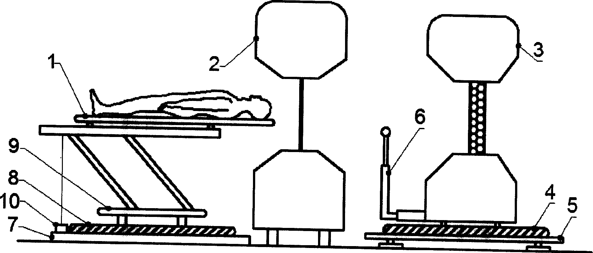
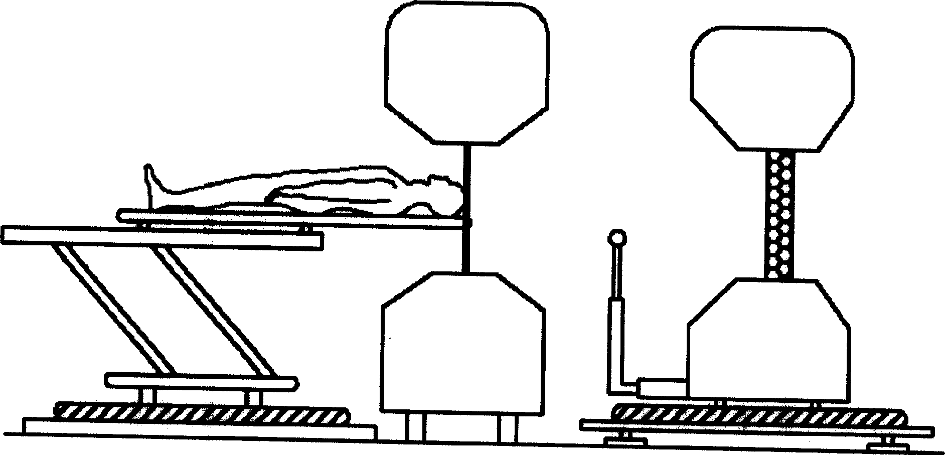
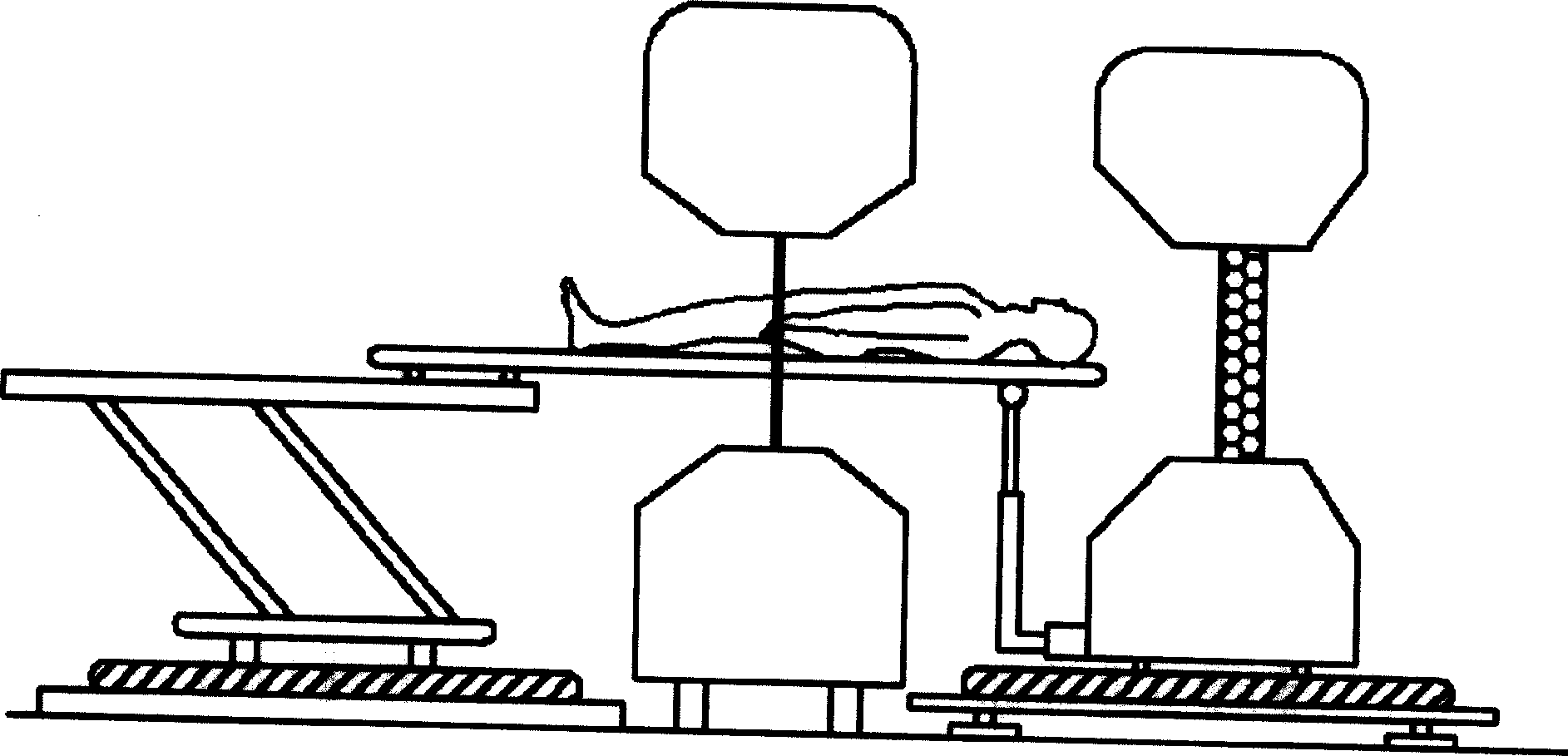
[0050] Such as figure 1 As shown, the multiple imaging system of the present invention includes a first imaging system 2, a second imaging system 3, a bed base 7, a guide rail II 8, a bed body 9, a bed board 1, and also includes a registration mechanism, which includes a guide rail I 4. Adjustable base 5, auxiliary support 6, displacement sensor 10, the first imaging system 2 is fixed on the horizontal ground, the bed body 9 is installed on the guide rail II 8, the bed body 9 can slide or roll along the guide rail II 8, and Can stay at any position on the guide rail. The guide rail II 8 is fixed on the bed base 7 with bolts, the bed base 7 is fixed on the ground, the second imaging system 3 is installed on the guide rail I 4, the second imaging system can slide or roll along the guide rail I 4, and can stay anywhere on the track. Guide rail I 4 is fixed on the adjustable base 5 with bolts, and the adjustable base 5 is placed on the ground; guide rail I and guide rail II ado...


PUM


Abstract

A multiple imaging system for precisely determining the position of lesion in human body is composed of the first imaging system, the second imaging system, a guide track, a bed body and its base, the bed board, and a regulation mechanism consisting of another guide track, regulatable base, auxiliary supporter, and displacement sensor.

Description

technical field [0001] The invention relates to the technical field of medical equipment, in particular to a multiple imaging system. Background technique [0002] In the field of medical imaging technology, according to the principle of imaging, it can be mainly divided into radiological imaging—that is, X-ray Transmission Computed Tomography (XCT), Magnetic Resonance Imaging (MRI) , Ultrasonography (Ultrasonography, USG) and radionuclide imaging - including gamma camera, single photon emission tomography (Single Photon Emission Computed Tomography, SPECT), positron emission tomography (Positron Emission Tomography, PET ). Among them, radiology imaging uses the difference in the absorption of X-rays by human tissues and organs, and converts analog information into photoelectric digital signals by detecting the remaining rays after penetrating the human body, and makes human tissues and organs into observable images through computer processing. Magnetic resonance imaging u...

Claims


Application Information

Patent Timeline
IPC IPC(8): A61B6/00A61B5/055
Inventor 郑全录高铁李甲递徐保伟于庆泽刘凯肖玲
Owner SHENYANG NEUSOFT PAISITONG MEDICAL SYST
Learn More