
Computer-tomographic system for carrying out a monitored intervention

A tomography and computer technology, which is used in radiological diagnostic instruments, computed tomography scanners, patient positioning for diagnosis, etc., can solve problems such as laboriousness, and achieve the effect of simple guidance and dose reduction

Inactive Publication Date: 2007-01-31
SIEMENS AG
View PDF0 Cites 7 Cited by
  • Summary
  • Abstract
  • Description
  • Claims
  • Application Information

AI Technical Summary

Problems solved by technology

With known CT patient couches, this control of the patient couches is relatively labor-intensive to carry out and poses relatively serious problems for the surgeon, especially when he is operating with sterile hands.
In addition there is a big problem when the instrument has to be introduced obliquely into the patient due to anatomy
Here it is particularly difficult to adjust the scan field to the actual intervention field

Method used


Image

  • Computer-tomographic system for carrying out a monitored intervention
  • Computer-tomographic system for carrying out a monitored intervention
  • Computer-tomographic system for carrying out a monitored intervention

Examples


Embodiment Construction

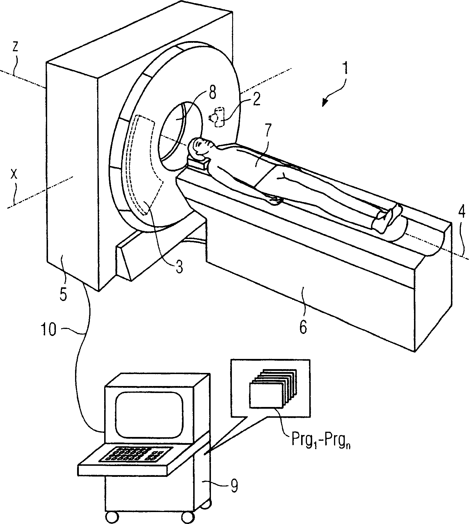
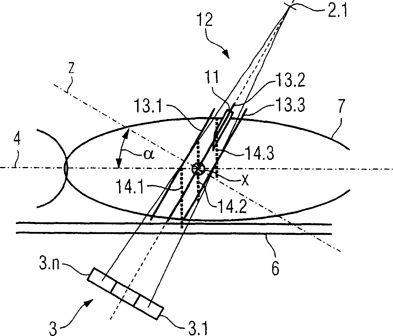
[0033] figure 1 A three-dimensional representation of a CT system 1 according to the invention is shown, which has a gantry housing 5 in which a gantry (not shown in detail) is arranged. The X-ray tube 2 and the detector 3 facing it are fixed on the bracket, which can rotate around the system axis 4 . The patient is situated on a patient couch 6 that is movable in the system axis direction 4 and can be moved through the opening 8 in order to be scanned in the radiation path in which the actual intervention is to be performed. Furthermore, a pivot axis x is shown perpendicular to the system axis z, around which the carrier can be pivoted so that the oblique radiation paths required by the invention can be realized. In this connection it should be noted that it is also within the scope of the invention for sufficiently wide, ie multi-line detectors, to shift only the calibration diaphragm so that only oblique rays are used for scanning.

[0034] Controlling of the reproduction...


PUM


Abstract

A CT system is disclosed for carrying out a monitored intervention with an instrument on a patient. The system includes an arrangement including an X-ray tube for production of a radiation beam which is moved around the patient and a detector with a large number of detector elements for measurement of the radiation intensity after passing through the patient, with the radiation beam scanning a scanning area on the patient. The system further includes an apparatus for variable orientation and positioning of the patient relative to the tube / detector arrangement, and a computation and control unit with computer programs or modules, for controlling the system and for reconstruction of the tomographic records from measurement data in the detector. A first program is stored for running and detects at least a portion of the instrument which is used for intervention, and a second program is stored for running and automatically matches the scanning area to the instrument, in accordance with presets that are provided.

Description

technical field [0001] The invention relates to a computed tomography system for performing an interventional intervention on a patient with an instrument, having at least one x-ray tube for generating a beam of radiation moving around the patient, and a detector comprising a plurality of detector elements, with After measuring the radiation intensity through a patient, wherein the radiation beam scans a scan area on the patient, the system also has means for variably supporting and positioning the patient relative to the x-ray tube / detector system, and includes a computer A computing and control unit of the program for controlling the system and reconstructing the tomographic image from the measurement data of the detector. Background technique [0002] In principle, such computed tomography systems are known, which are usually used for invasive interventions on patients under simultaneous control. Such invasive interventions are, for example, extraction of tissue or treat...

Claims


Application Information

Patent Timeline
Patent Type & Authority Applications(China)
IPC IPC(8): A61B6/03A61B6/04
CPCA61B6/12A61B6/032A61B6/587A61B6/02A61B6/506
Inventor 加布里埃尔·哈拉斯
Owner SIEMENS AG
Learn More