Oral cavity image diagnostic device

An image diagnosis and imaging device technology, which is applied in diagnosis, dental mirror, diagnosis recording/measurement, etc., can solve the problems of image framing, unfreezing, inconvenient use, and affecting diagnosis results, etc., to achieve fast and flexible access , Improve efficiency, facilitate recording and retrieval

Inactive Publication Date: 2007-03-14
西安北方光电有限公司
View PDF0 Cites 2 Cited by
  • Summary
  • Abstract
  • Description
  • Claims
  • Application Information

AI Technical Summary

Problems solved by technology

[0004] This technology can observe and wipe the pathological conditions inside the oral cavity through dynamic images, but there are still problems of inconvenient use, such as the frame fixing and thawing of images cannot be completed conveniently, which affects the diagnosis results

Method used


Image

  • Oral cavity image diagnostic device
  • Oral cavity image diagnostic device

Examples


Embodiment Construction

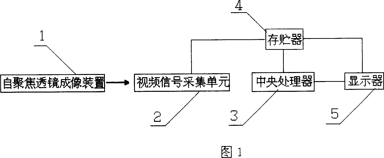
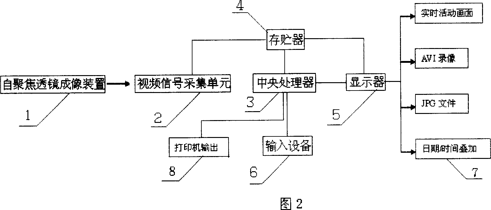
[0020] As shown in Figure 2, oral cavity image diagnostic instrument device, it comprises: self-focusing lens imaging device 1, is used for oral cavity image imaging on the CCD device target surface; Provides video signal acquisition unit 2; Video signal acquisition unit 2, uses The video signal output by the CCD device is sampled, held, and A / D converted; the imaging information used for A / D conversion is stored in the memory 4; the imaging information is input to the central processing unit CPU 3 at the same time; the central processing Processor CPU 3 digitally processes and compresses the imaging information of the A / D conversion, generates moving and static pictures respectively, and stores them into corresponding file forms and inputs the above-mentioned moving and static pictures to the display 5 for display; The device 6 selects the above-mentioned active and static pictures for display.

[0021] The dental image diagnostic instrument device can be explained better thr...


PUM


Abstract

The invention pertains to iatrical equipment, particularly the diagnostic apparatus for images of nonnasality. Its features are : auto-focus lens imaging devices for imaging the oral images on the CCD target surface; video signal acquisition unit for sampling, maintaining and A / D transforming the video signal out from the CCD, for storing the imaging information out from the A / D transformation, and for inputting the stored image data to CPU; the CPU for processing in digital way and compressing the imaging information out from A / D conversion, bringing out static and active pictures respectively and stored into the documentation of corresponding form, and for inputting the said active and static pictures into the monitor ; monitor, for displaying the said active and static images; input devices, for choosing the said active or static images to display. Surgeons and patients both distinctly observe the images. The invention makes the communication between each other easier and improves the success rate and the efficiency of surgery effectively.

Description

technical field [0001] The invention belongs to medical equipment, in particular to an oral image diagnostic instrument device. Background technique [0002] With the help of computer and video technology, the measured object (object image) is imaged on the target surface of a special CCD device by means of self-focusing lens imaging technology, and the generated video signal is processed by sampling and holding compression, and finally displayed on the computer monitor. Use this method to scan the pathological conditions inside the oral cavity, and process and process the dynamic image information through the specific control mode of the man-machine interface to achieve the purpose of application. [0003] As shown in Figure 1, the object to be measured is imaged on the CCD device target surface (probe) by a focusing lens to form a self-focusing lens imaging device 1, and the video signal output by the self-focusing lens imaging device 1 is sampled through a video signal ac...

Claims


Application Information

Patent Timeline
Patent Type & Authority Applications(China)
IPC IPC(8): A61B5/00A61B1/24
Inventor 彭纪辉马利民曹晓玲
Owner 西安北方光电有限公司
Why Eureka
Try Eureka