Immunodiagnosis reagent kit for breast cancer and testing sheet
A diagnostic kit and immunohistochemical technology, applied in the field of immunology, can solve the problems of cumbersome operating procedures and high non-specificity
- Summary
- Abstract
- Description
- Claims
- Application Information
AI Technical Summary
Problems solved by technology
Method used
Image
Examples
Embodiment 1
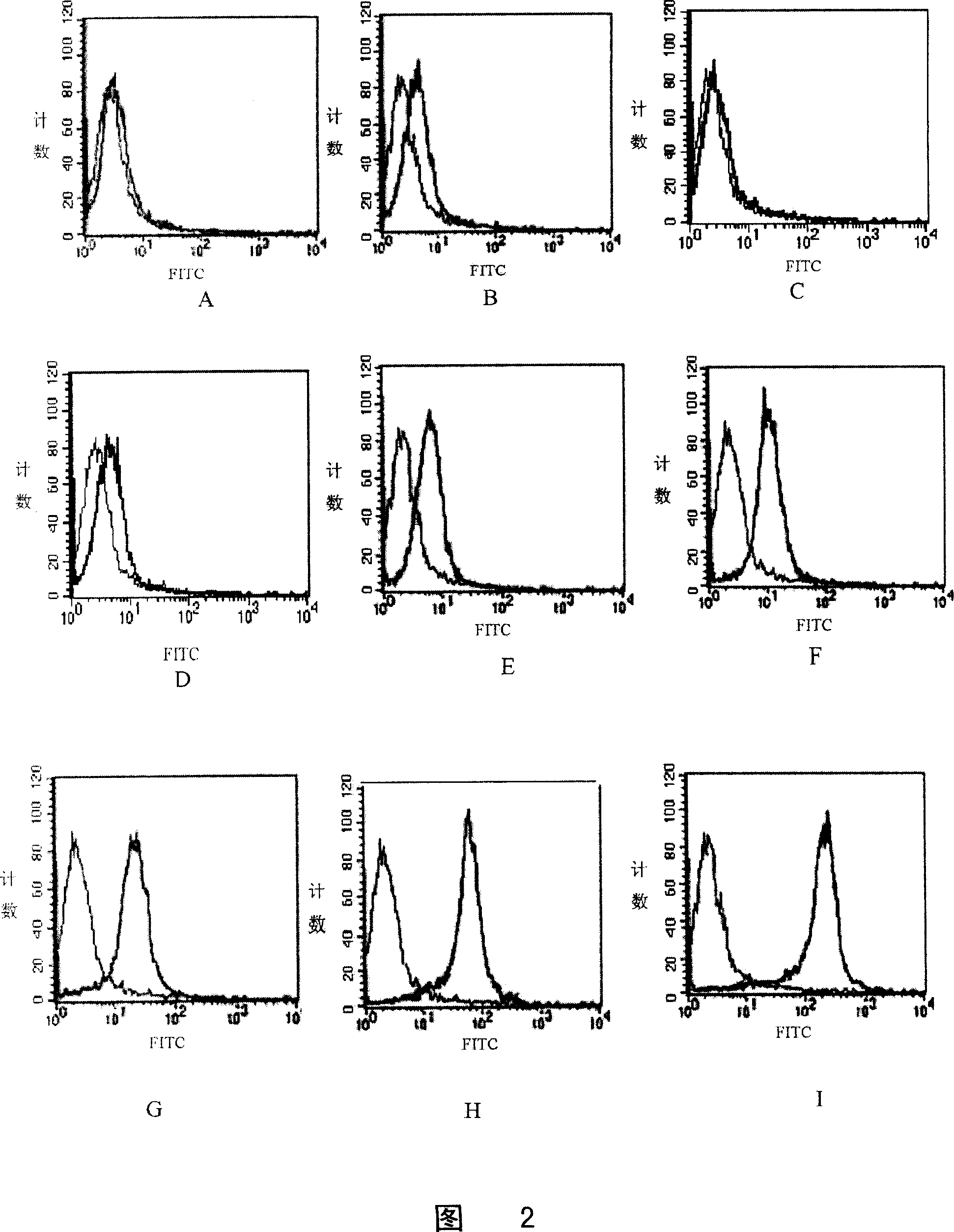
[0039] Example 1 Selection of tumor cells with different expression levels of Her2 antigen
[0040] Tumor cell selection was performed using the following steps:
[0041] 1) Alternative tumor cells include: human liver cancer cells QYC, human liver cancer cells KYC, human liver cancer cells LYX, mouse renal adenocarcinoma cells RENCA, D2F2 / E2 (the above five cells were obtained from the International Cooperation Tumor Research Institute of Second Military Medical University ), human epidermal cancer cell A431 (purchased from Shanghai Wuli Biotechnology Co., Ltd.) and MCF-7 (obtained from the National Cancer Institute of the United States), SK-BR-3 (purchased from Shanghai Wuli Biotechnology Co., Ltd.), HTB -20 (purchased from Guangzhou Wellman Company). After culturing in vitro to the logarithmic growth phase, various cells were taken for fluorescent staining;
[0042] 2) For cell counting, add the tumor cells to be tested into a special detection tube for flow cytometry, 2×...
Embodiment 2
[0049] Example 2 Preparation of Tumor Tissue Expressing Her2 Antigen Homogeneity
[0050] Preparation:
[0051] 1) Three tumor cells, D2F2 / E2, LYX and MCF-7, which were obtained in Example 1 and showed high, moderate and no expression of Her2 respectively, were selected and passaged in RPMI-1640 complete medium containing 10% fetal bovine serum Expansion, culture conditions are 5% CO 2 , 37°C and saturated humidity;
[0052] 2) For the three types of cells in the logarithmic growth phase, wash with PBS, centrifuge at 200g for 5 minutes, discard the supernatant, resuspend the cell pellet in PBS, count, and adjust the cell density to 1×10 7 / mL;
[0053] 3) The tumor cell suspension was inoculated subcutaneously in the neck of Balb / c nude mice respectively, and the local tumor growth and the general condition of the tested animals were regularly observed;
[0054] 4) When the tumor grows to 0.5cm 3 Left and right size (the formula for calculating tumor volume is x 2 y / 2, w...
Embodiment 3
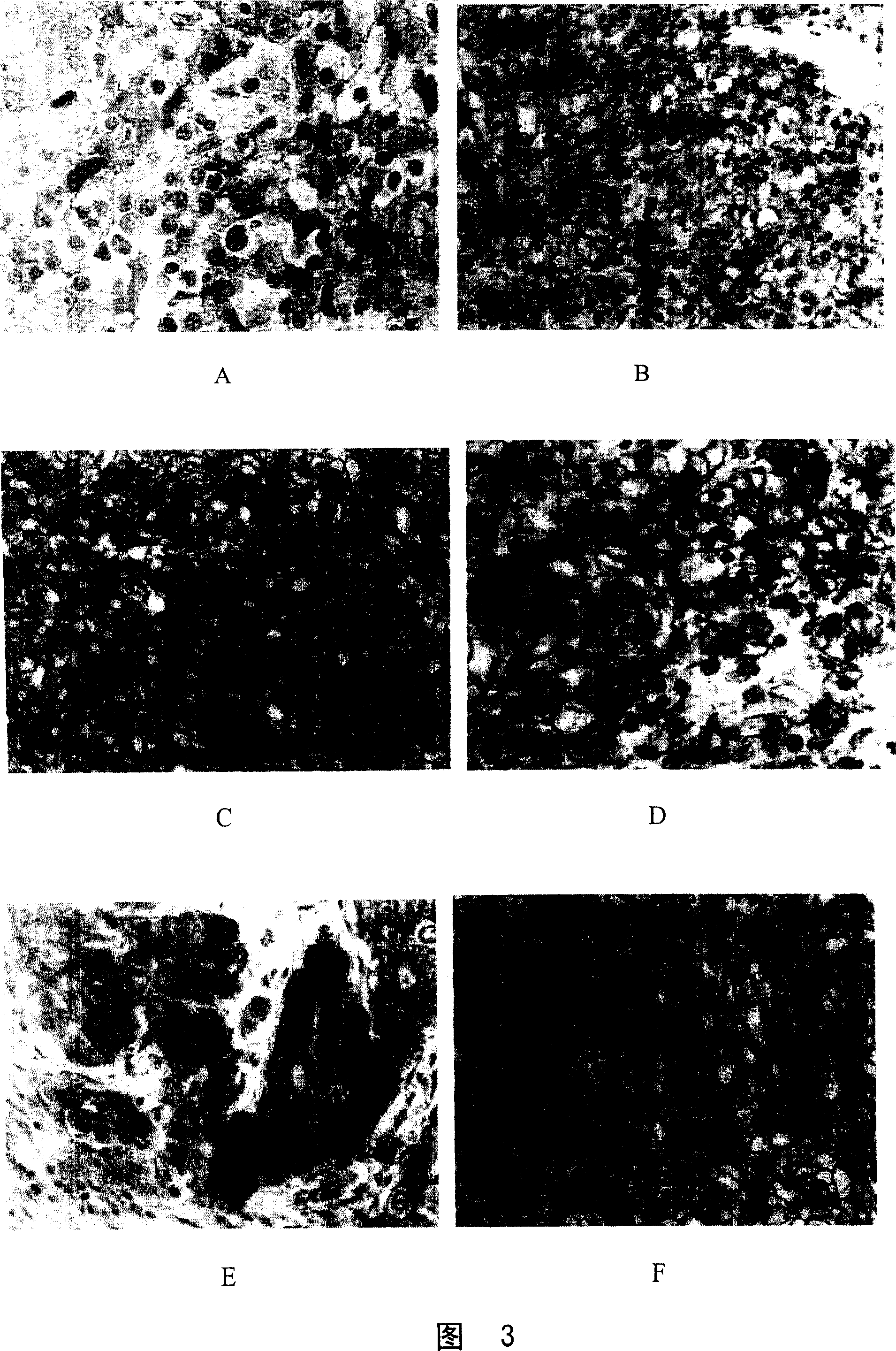
[0061] Example 3 Preparation of Tumor Histopathological Sections and Detection of Surface Her2 Antigen Expression
[0062] 1. Production
[0063] 1) Preparation of paraformaldehyde phosphate buffer solution: 40g of paraformaldehyde, 500mL of 0.1mol / L PBS with pH7.4, mix the two and heat to 60°C, stir and add 1N NaOH dropwise until the solution is clear, then add PBS to The total volume is 1000 mL.
[0064] 2) After removing the tumor tissue, process the tissue as soon as possible, and try to keep the tissue fresh and not dry, and the tissue block should not be too large or too thick, especially the thickness should not exceed 1cm.
[0065] 3) The collected tissue is fixed in paraformaldehyde phosphate buffer solution. The amount of fixative solution should be sufficient, at least 20 times that of the tissue. Otherwise, the concentration of the fixative solution will decrease. Too long time may affect the dyeing effect.
[0066] 4) Dehydration, transparency, and wax-immersio...
PUM
 Abstract
Description
Claims
Application Information
 - R&D
- Intellectual Property
- Life Sciences
- Materials
- Tech Scout
- Unparalleled Data Quality
- Higher Quality Content
- 60% Fewer Hallucinations
Browse by: Latest US Patents, China's latest patents, Technical Efficacy Thesaurus, Application Domain, Technology Topic, Popular Technical Reports.
© 2025 PatSnap. All rights reserved.Legal|Privacy policy|Modern Slavery Act Transparency Statement|Sitemap|About US| Contact US: help@patsnap.com


