Method and system for tomographic imaging using fluorescent proteins

A fluorescent protein, optical tomography technology, used in diagnosis, application, diagnostic recording/measurement, etc., to solve problems such as limiting the quality of final optical tomography images

Inactive Publication Date: 2007-04-25
THE GENERAL HOSPITAL CORP
View PDF0 Cites 15 Cited by
  • Summary
  • Abstract
  • Description
  • Claims
  • Application Information

AI Technical Summary

Problems solved by technology

Room temperature or moderately cooled CCD cameras are known to have high levels of ...

Method used


Image

  • Method and system for tomographic imaging using fluorescent proteins
  • Method and system for tomographic imaging using fluorescent proteins
  • Method and system for tomographic imaging using fluorescent proteins

Examples


Embodiment Construction

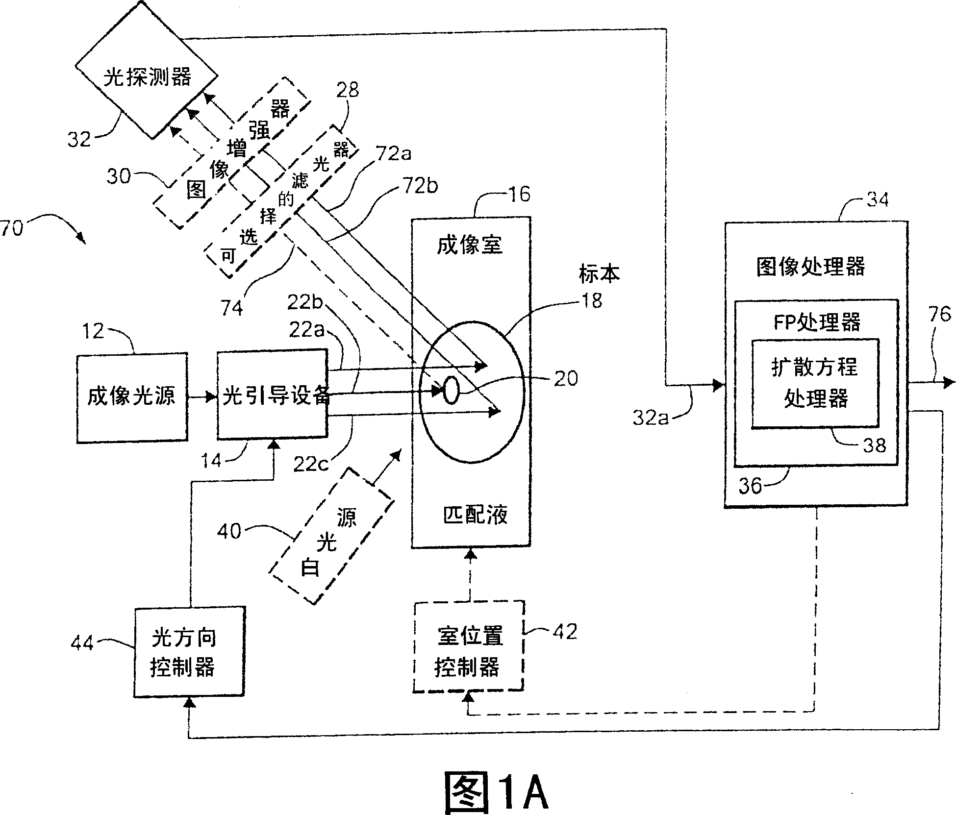
[0035] Before introducing the imaging methods and systems, some introductory concepts and terminology are explained. As used herein, "phantom" refers to the test object being imaged. The phantom is typically an artifact, such as a piece of plastic, with diffuse light propagation properties similar to living tissue. As another example, the phantom can be a glass vial with cells expressing a fluorescent protein (ie, fluorescently labeled) therein.

[0036] As used herein, the term "apparent light source" is used to describe a single light source projected to multiple physical positions or angles, each providing an apparent light source.

[0037] As used herein, the term "excitation" light is used to describe light produced by an excitation source, such as an apparent source, which travels toward a specimen to be imaged before entering the specimen. Once in the specimen, the light is referred to herein as "intrinsic" light. This intrinsic light is absorbed and scattered in the...


PUM


Abstract

A system for optical tomography includes an apparent light source adapted to project excitation light toward a specimen having fluorescent proteins therein, wherein the excitation light enters the specimen becoming intrinsic light within the specimen, wherein the intrinsic light is adapted to excite fluorescent light from the fluorescent proteins, and wherein the intrinsic light and the fluorescent light are diffuse. A method of optical tomography includes generating the excitation light with the apparent light source, wherein the intrinsic light and the fluorescent light are diffuse.

Description

technical field [0001] The present invention relates generally to optical tomography, and more particularly to methods and systems for extracting quantitative three-dimensional molecular and biological information from living specimens using fluorescent proteins. Background technique [0002] Fluorescent proteins (FPs) are important reporters for different biomedical applications. In some existing applications, engineered FPs are probed using epifluorescence, confocal (microscopy) or reflectance (whole animal) imaging. [0003] Epi-fluorescence, confocal microscopy, depends on coherent (non-diffusing) light projected onto and reflected from the specimen. Because microscopy requires intrinsically coherent light, this technique can only image small depths (eg, less than 1 mm) into a specimen. At deeper imaging depths, light is known to start to diffuse, making microscopy ineffective at deeper imaging depths. [0004] Reflected fluorescence imaging has been shown to be usefu...

Claims


Application Information

Patent Timeline
IPC IPC(8): A61B5/00
Inventor 瓦西利斯·恩齐亚克里斯托斯豪尔赫·里波利扬尼斯·扎哈拉基斯
Owner THE GENERAL HOSPITAL CORP
Why Eureka
Try Eureka