Accelerated data capture in an x-ray system

a data capture and x-ray technology, applied in the field of x-ray mammography, can solve the problems of reducing the number of available information about objects, limiting the use of tomosynthesis, and the data capturing time of the ffdm, so as to reduce the reading time, and reduce the number of reads

Active Publication Date: 2018-07-24
SIEMENS HEALTHCARE GMBH
View PDF5 Cites 1 Cited by
  • Summary
  • Abstract
  • Description
  • Claims
  • Application Information

AI Technical Summary

Benefits of technology

[0008]The fact that only some of the pixels of the detector for capturing the data are evaluated provides that the reading time of the detector may be advantageously shortened. As a result, the time period for detecting the data may be shortened in comparison with the prior art, without reducing the number of projections, and consequently the image quality.
[0038]In the case of mammography, the reduction of the reading time according to one or more of the present embodiments allows a tomosynthesis scan to be carried out just as quickly as a two-dimensional screening. The fact that the set of data read is reduced in comparison with the prior art provides that the transmission time of the data and the storage space for storing the data may also be advantageously reduced.

Problems solved by technology

The data capturing time of the FFDM, and consequently the reading time for each projection image of the FFDM, is currently a very limiting factor with respect to the use of tomosynthesis.
However, this approach has the great disadvantage that the available information about the object decreases with the number of projections captured.

Method used


Image

  • Accelerated data capture in an x-ray system
  • Accelerated data capture in an x-ray system
  • Accelerated data capture in an x-ray system

Examples


Embodiment Construction

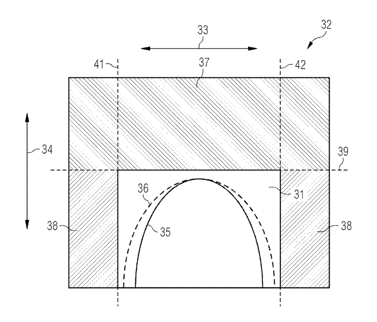
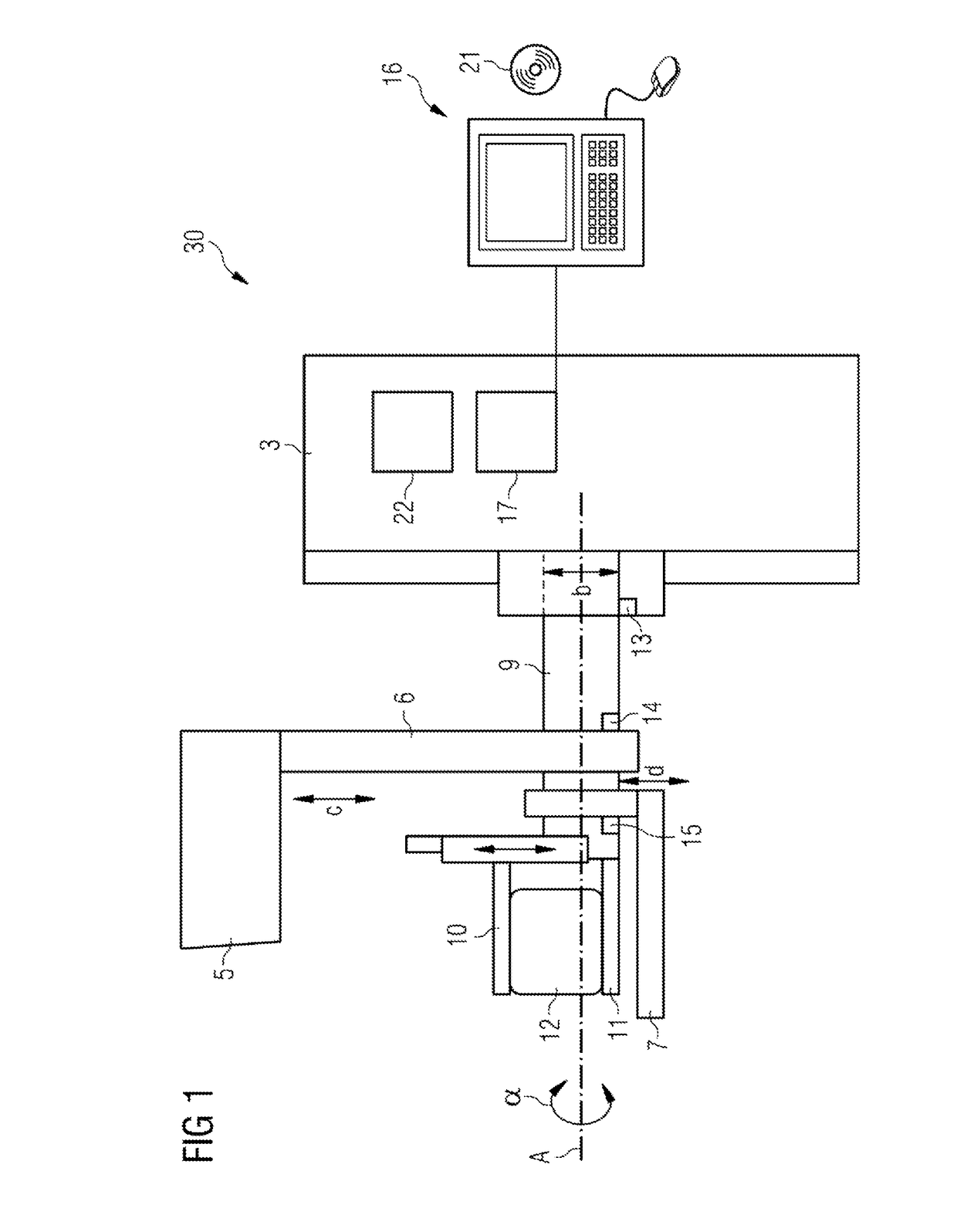
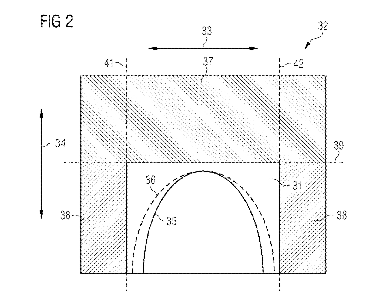
[0042]In FIG. 1, an x-ray system 30 for mammography examinations according to one or more of the present embodiments, which may also be referred to as a tomosynthesis machine, is schematically represented. The x-ray system 30 includes a carrying arm 9 that is mounted pivotably about a horizontally running axis A in a mounting (compare double-headed arrow or angle α). The mounting is arranged on a stand 3 and is vertically adjustable, as indicated by the double-headed arrow b. Arranged on the carrying arm 9 are an arm 6 provided with an x-ray source 5, an area detector 7 (e.g., FFDM) and a compression device including a compression plate 10 and a bearing plate 11. In FIG. 1, a female breast 12 compressed by the compression plate 10 and the bearing plate 11 is represented in a schematic way. The arm 6 is pivotable about the axis A in relation to the carrying arm 9, the detector 7 and the compression device 10, 11. For height adjustments and pivoting movements, electric motors 13 to 15...


PUM


Abstract

Data of a predetermined volume portion of an object under examination is captured by an x-ray system that includes an x-ray source and a detector. The x-ray source is activated to generate x-rays that emerge from the x-ray source, radiate through the volume portion, and after radiating through, impinge on the detector. X-rays impinging on the detector are captured pixel by pixel, in order to capture the data of the predetermined volume portion. With the pixel-by-pixel capture, only a subset of all the pixels of the detector is evaluated.

Description

[0001]This application claims the benefit of DE 10 2014 224 743.8, filed on Dec. 3, 2014, which is hereby incorporated by reference in its entirety.BACKGROUND[0002]The present embodiments relate to accelerated data capture.[0003]In the area of x-ray mammography, the significance of tomosynthesis has increased in recent years. In tomosynthesis, x-ray images are captured over a limited angular range. The projection images thereby determined may be reconstructed in the form of tomograms of any desired alignment. Often used as the x-ray detector for this is a flat field detector for mammography (FFDM) that represents a key component for the tomosynthesis. The data capturing time of the FFDM, and consequently the reading time for each projection image of the FFDM, is currently a very limiting factor with respect to the use of tomosynthesis.[0004]In order to shorten the overall data capturing time in tomosynthesis, currently the number of projections captured is restricted. However, this ...

Claims


Application Information

Patent Timeline
Patent Type & Authority Patents(United States)
IPC IPC(8): G06K9/00A61B6/03A61B6/00A61B6/02
CPCA61B6/5205A61B6/025A61B6/4233A61B6/58A61B6/54A61B6/542A61B6/502
Inventor BARTL, PETERJEREBKO, ANNAWEIDNER, TOM
Owner SIEMENS HEALTHCARE GMBH
Why Eureka
Try Eureka