Device for producing cuts or perforations on an eye

a technology for producing eye cuts and perforations, applied in the field of eye cuts or perforations, can solve the problems of affecting vision, affecting the ability of the eye to heal, and affecting the effect of vision,

Inactive Publication Date: 2019-02-26
VOSSAMED
View PDF13 Cites 0 Cited by
  • Summary
  • Abstract
  • Description
  • Claims
  • Application Information

AI Technical Summary

Benefits of technology

[0017]It is possible for the cutting element—unlike known instruments—to have no mechanical connection to a handle held by the surgeon or the like, and in addition to being merely set into rotational or oscillatory motion by means of the (electro)magnetic field generated by the field generator, is also held in its desired position in the eye where the surgeon has previously brought it. This means that not only can the cutting element be held in the desired centered position with the field generator, but it is also possible to exert force on the cutting element in the axial direction, to that it is held pressed against the capsule to be cut, and consequently the cut is effected around the entire circumference in a particularly easy and reliable manner.
[0018]One embodiment in which the cutting element mounted on a bearing in a holding element such that is free to move, in particular to rotate, and with which it can be introduced into the interior of the eye through an opening, has proved to be especially advantageous. With the aid of the holding element, the surgeon, after he has opened up the anterior chamber of the eye with a small cut or hole, can introduce the cutting element, which is mounted in bearing at the remote, free end of the holding element, into the eye and hold it in the desired position while the (rotary) drive of the cutting element then takes place without any contact by switching on the magnetic field generated by the field generator. The holding element can of course also be manipulated for this purpose by a robot provided for this purpose that can handle the tool with high precision and hold it reliably in the found desired position.
[0028]The invention therefore creates a new device for perforating / cutting tissue, particularly in or at the eye. The device is generally used for anterior and posterior perforation (capsulotomy, rhexis) of the human eye lens. The device comprises a cutting element or cutting head that can be introduced into the eye through a small incision and has a device arranged outside the eye with which mechanical vibrations or movements are produced by electromechanical means, as if with a small electric motor, at the cutting element. The cutting element set into mechanical, mostly oscillating, vibrations in this way produces a cutting effect in the eye due to its direct contact with the tissue, without the cutting element having to be coupled mechanically to an external drive through the small incision in the eye, and in one preferred embodiment it is not even necessary for the cutting element to held from outside by a holder the cutting process. The opening of the eye lens capsule has a diameter in the range of a few tens of μm to a few millimeters (e.g. 0.01 mm to 10 mm).
[0031]The frequency of the vibrations is however chosen so that a clean perforation of the lens capsule is obtained, which as a rule is ensured if the frequency is either substantially smaller or greater than the natural frequency of the tissue to be cut. Ideally, it lies in the range from a few Hz through the kHz range into the MHz range (ultrasound). The vibration of the cutting element or a cutting edge mounted on it is only necessary for a short time (<1 minute) to perform the necessary cut,

Problems solved by technology

If this stability / integrity of the rhexis is not ensured, tears can arise in the capsule sac which can result in complications during the operation (e.g. loss of lens material into the vitreous humour space, difficulty to impossibility of implanting an artificial intraocular lens IOL, prolapse of the vitreous humour, etc.).
The so-called “secondary cataract” can lead to distortion of the capsule sac and clouding of the remaining parts of the capsule.
The posterior lens membrane can become grown over with epithelium cells, become cloudy and severely impair the vision.
Too small a rhexis can lead to subsequent complications (capsular phimosis) in just the same way as too large a rhexis compared to the IOL (OIL lens becoming caught in the rhexis, and off-centerdness of the IOL).
A rhexis which is off-center from the IOL, which does not cover the front of the IOL in a circular manner, can lead to pressure by the shrinking rhexis edge on the IOL lens and to the IOL being off-center.
A posterior rhexis is technically more difficult to accomplish, however, and more risky from a medical aspect.
However, these methods still have the disadvantage, among others, that all they can do is produce a point-shaped perforation at a particular place.
This has the disadvantage of introducing electric current into the eye and also heating up the ocular tissue.
The expansion of the cavitation bubble this causes finally tears the tissue locally.
However, the patient's eye has to be firmly connected to such a laser device for this.
This prolongs the operation time and procedure.
In addition, the devices are very large, demand considerable maintenance, and are expensive.
These have a number of disadvantages: Attaching to the lens capsule by suction within the eye is not without risks, since the liquid reservoir in the anterior chamber of the eye is limited and the anterior chamber can therefore collapse, thereby damaging the ocular tissue.
This is particularly likely because the suction device in the eye has to operate with the corresponding vacuum, and must therefore be quite large and non-portable.
The instrument is relatively bulky and not easy for the surgeon to handle.
Both known instruments share a common feature in that they need to be held and manipulated by the surgeon on the handle or shaft respectively during the cutting process, which means that every incorrect manipulation inevitably affects the cut to be made.
This has the disadvantage that the drive energy for cutting element has to be transmitted into the eye by means of moving parts, i.e. the drive and cutting element are joined together mechanically.

Method used


Image

  • Device for producing cuts or perforations on an eye
  • Device for producing cuts or perforations on an eye
  • Device for producing cuts or perforations on an eye

Examples


first embodiment

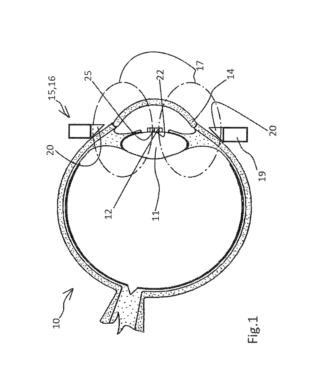
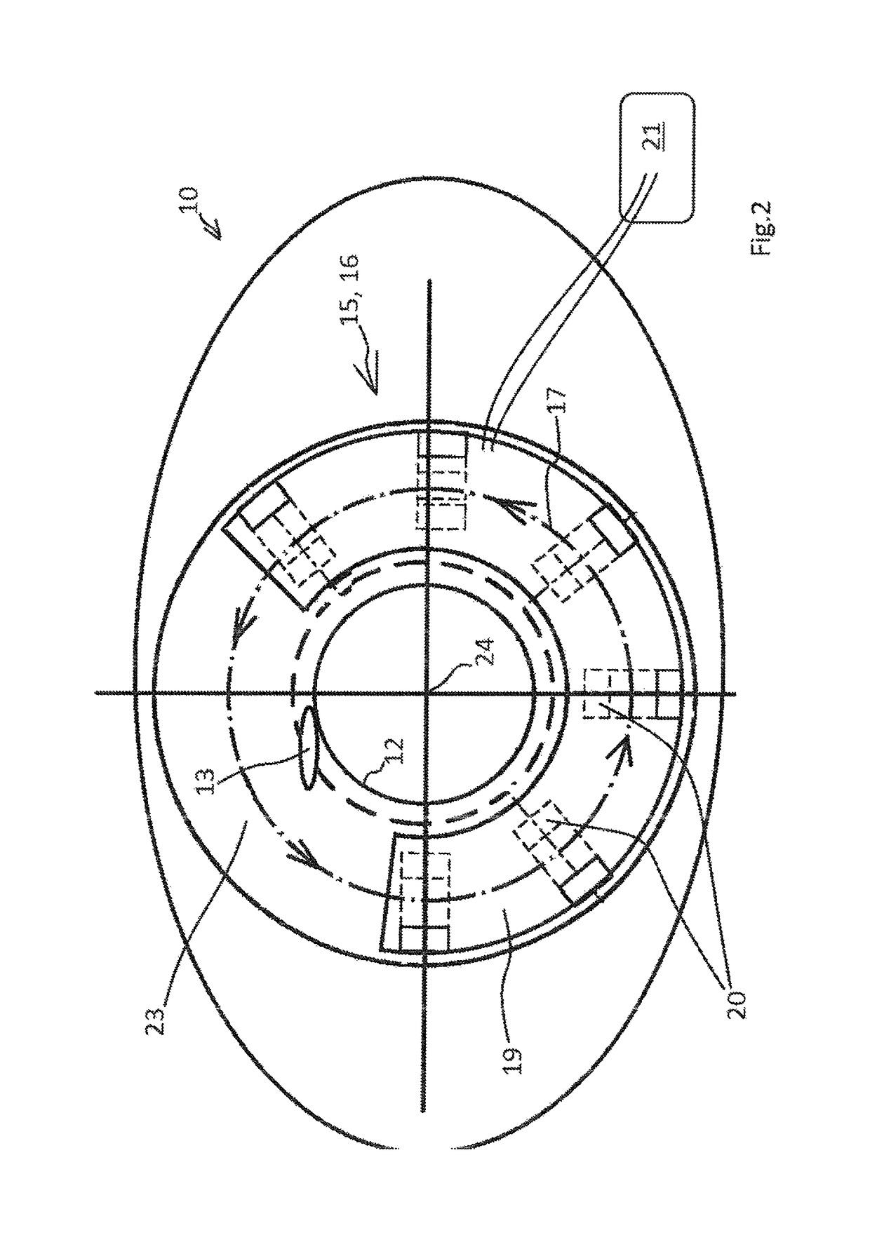
[0041]Referring now to the drawings wherein the showings are for the purpose of illustrating preferred and alternative embodiments of the invention only and not for the purpose of limiting the same, FIGS. 1 and 2 show a device according to the invention used for producing a rhexis, in other words a circular cut in the eye 10 for opening the eye lens 11. The device includes here a cutting element 12, which by means of a small incision 13 in the cornea 14 of the eye 10 to be operated on can be introduced into the interior of the eye. The cutting element can be elastically deformable for this, so as to permit to be introduced, similar to an intraocular lens, into the interior of the eye through the comparatively small cut 13 using methods familiar to the eye surgeon. The cutting element is magnetisable or magnetic, for which purpose it is either completely made of metal with ferromagnetic properties, or has a metallic coating, a ferromagnetic core or a bracing made of (ferromagnetic) m...

second embodiment

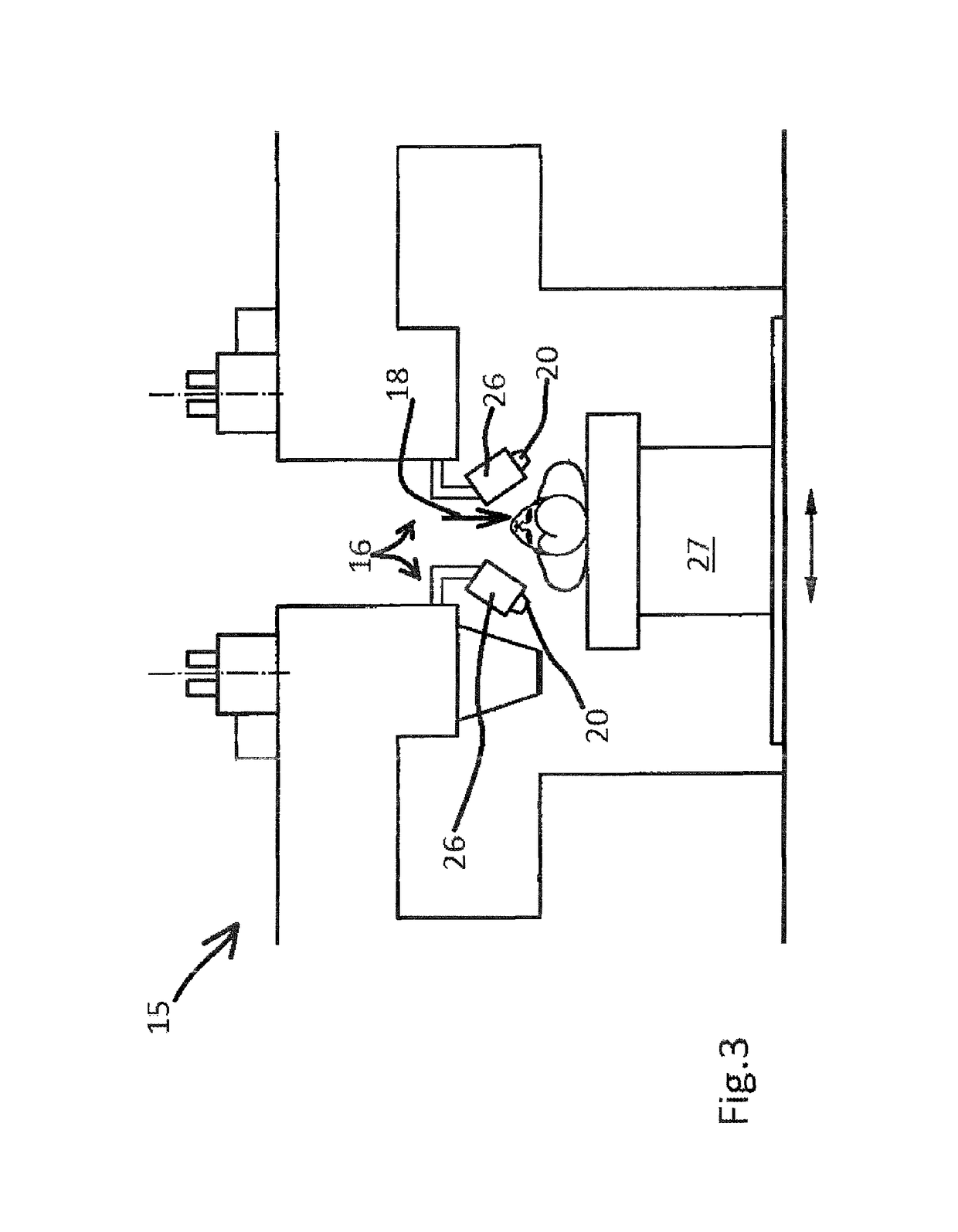
[0046]The second embodiment illustrated in FIG. 3 differs from the embodiments in FIGS. 1 and 2 mainly in that the field generator for generating the electromagnetic field is not arranged here in the immediate vicinity of the cutting element directly at the eye of the patient to be operated upon, but on an item of equipment that surrounds the entire head of the patient at a greater distance from the cutting element. The exciter magnets here are arranged on magnet holders 26, of which there are several, preferably arranged at even angular distances, surrounding the head of patient at the height of eye to be operated upon. It can be seen in the embodiment shown in FIG. 3 that the operating table 27 with the patient lying on it can be moved sideways so as to adjust the eye to the optimal position relative to the magnet holders 26.

[0047]FIGS. 4 and 5 show and alternative design of a cutting element used in the invention. In this embodiment, the cutting element 12 has two cloverleaf-shap...


PUM


Abstract

A Device for producing cuts or perforations on a human or animal eye at the anterior or posterior opening of the eye lens (capsulatomy, rhexis) with a cutting element that can be inserted into the interior of the eye and a drive device which is arranged outside of the eye in order to cause movement, in particular oscillatory vibrations or rotation of the cutting element inside the interior of the eye, the drive device having a field generator for generating a magnetic or electromagnetic excitation field and the cutting element can be excited, in particular driven, by way of the excitation field generated by the field generator.

Description

[0001]The invention refers to a device for producing cuts or perforations on a human or animal eye at the anterior or posterior opening of the eye lens (capsulatomy, rhexis) with a cutting element that can be inserted into the interior of the eye and a drive device which is arranged outside of the eye in order to cause movement, in particular oscillatory vibrations or rotation of the cutting element inside the interior of the eye. The device according to the invention is therefore particularly suitable for use in ophthalmic surgery, especially for cutting out parts of the capsule of the human eye lens.BACKGROUND OF THE INVENTION[0002]Operations on the human or animal eye lens belong to the operations most frequently carried out worldwide. Such operations become necessary as a result of pathological processes which mostly affect the interior of the eye lens, such as clouding of the eye lens (cataract formation) and / or hardening of the lens nucleus in presbyopia or if a change in the ...

Claims


Application Information

Patent Timeline
Patent Type & Authority Patents(United States)
IPC IPC(8): A61F9/007
CPCA61F9/00754
Inventor GERTEN, GEORG
Owner VOSSAMED
Why Eureka
Try Eureka