

Acquisition and processing of measurement data by a combined magnetic resonance and X-ray device
a combined magnetic resonance and x-ray technology, applied in the field of acquisition and processing of measurement data by a combined magnetic resonance and x-ray device, can solve the problems of reducing image quality and image artefacts, unable to track an internal movement of the test subject, and the three-dimensional image acquisition in magnetic resonance imaging takes a certain amount of time, so as to achieve the effect of improving the measurement quality
- Summary
- Abstract
- Description
- Claims
- Application Information
AI Technical Summary
Benefits of technology
Problems solved by technology
Method used
Image

Examples
Embodiment Construction
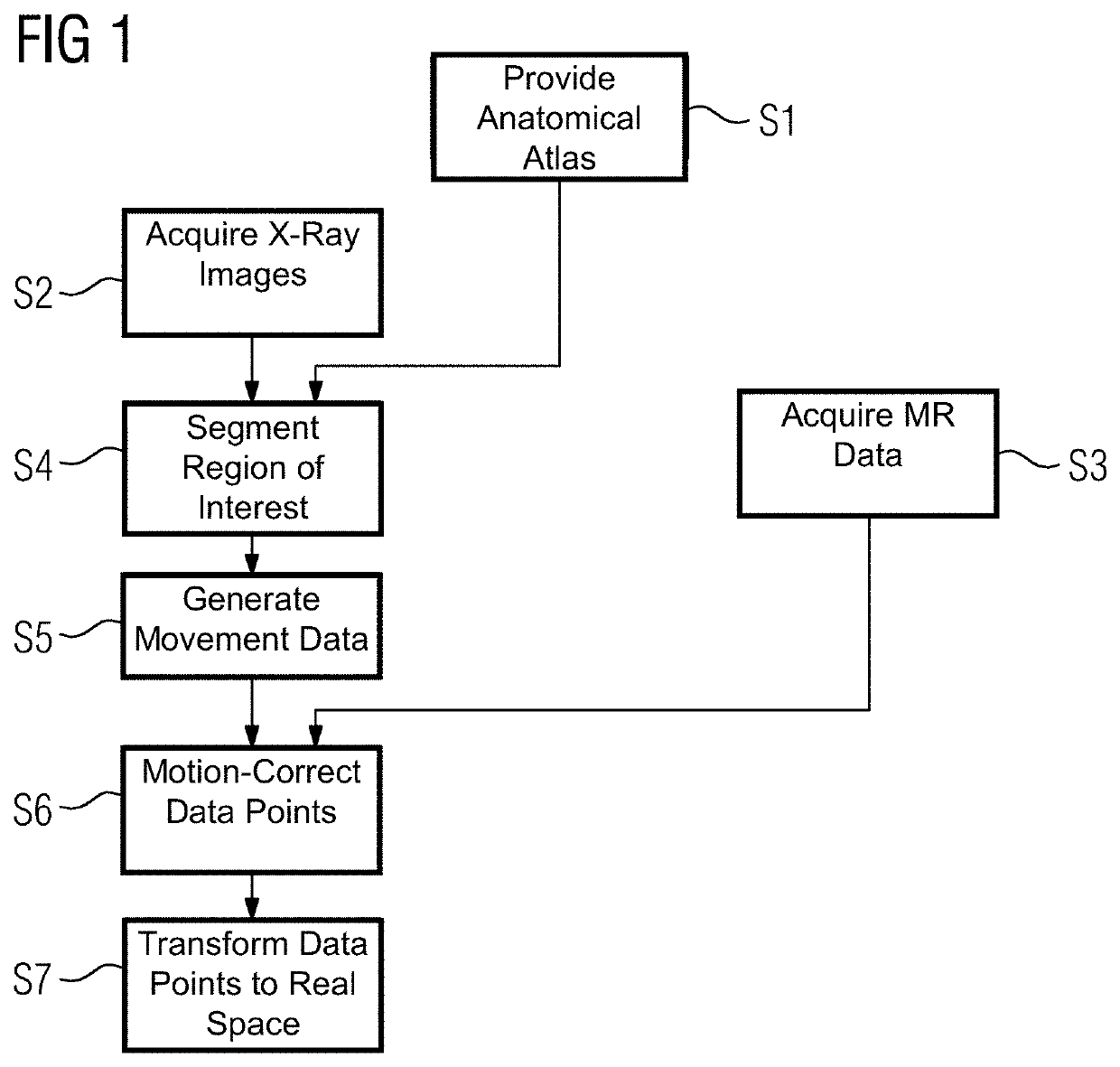
[0034]FIG. 1 shows one embodiment of a method for the acquisition and a processing of measurement data by a combined magnetic resonance and X-ray device. In act S1, prior knowledge about the test subject is provided in the form of an anatomical atlas that will be used later in the method to determine relevant regions (e.g., a heart), for which a movement correction may be provided.
[0035]In act S2, a plurality of X-ray images are acquired in succession by a X-ray acquisition unit. During the same time interval, in which the X-ray images are acquired, several data points representing a magnetic resonance signal strength for different phase encodings are acquired by a magnetic resonance acquisition unit during act S3. The acquisition of the different data points is done according to the prior art in magnetic resonance imaging. A certain slice of the test subject is excited by an excitation pulse, a phase encoding gradient is applied to provide a phase encoding, and the radiation due to...
PUM
 Abstract
Description
Claims
Application Information