
Multi-chain polypeptide-containing tri-specific binding molecules that specifically bind to multiple cancer antigens
a tri-specific binding and antigen technology, applied in the field of tri-specific binding molecules, can solve the problems of limiting the duration and effectiveness of human therapeutic applications of those moities, no mechanism for their production, and compromises and trade-offs, and achieve the effect of facilitating the killing of such cancer cells
- Summary
- Abstract
- Description
- Claims
- Application Information
AI Technical Summary
Benefits of technology
Problems solved by technology
Method used
Image

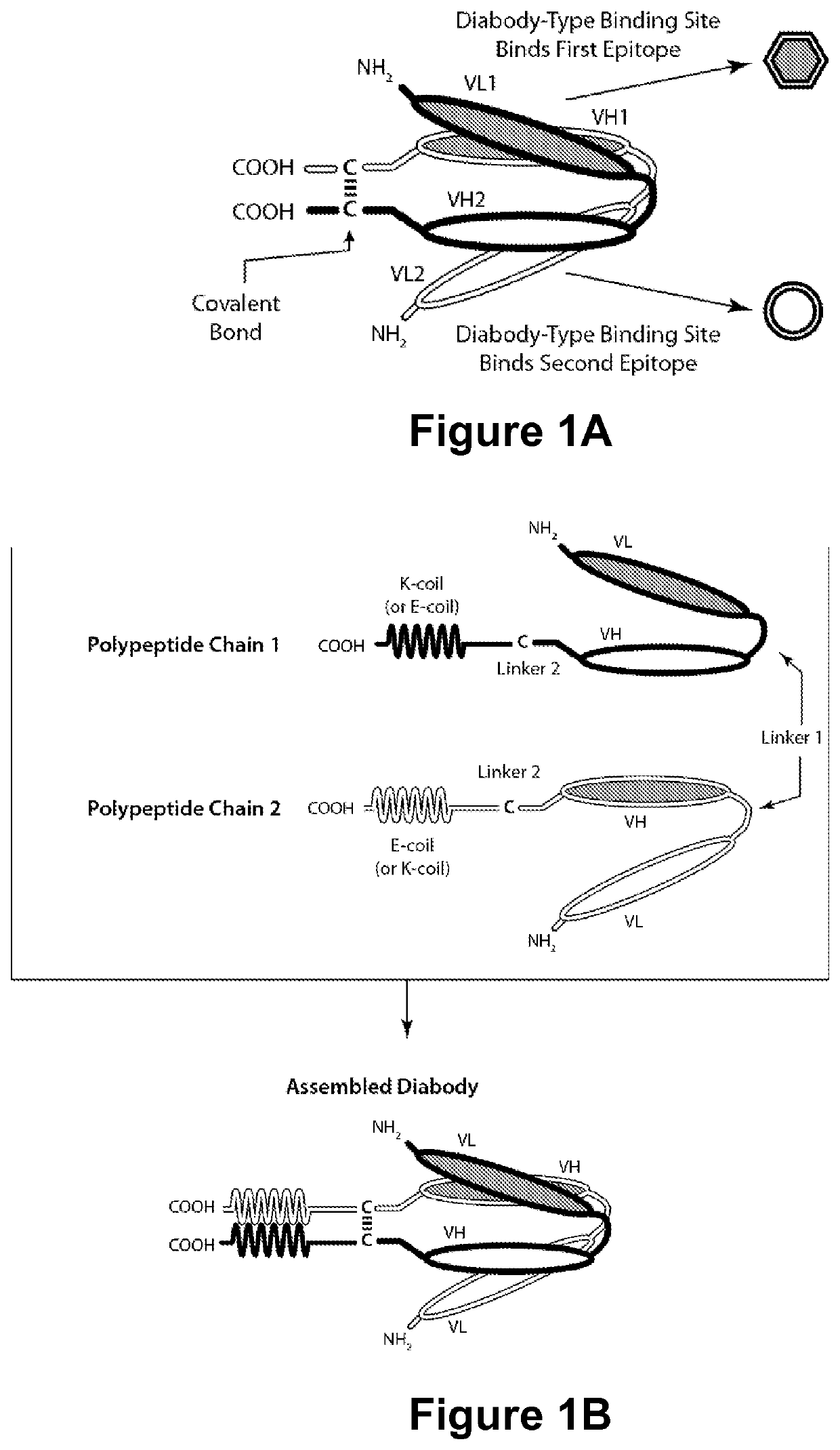
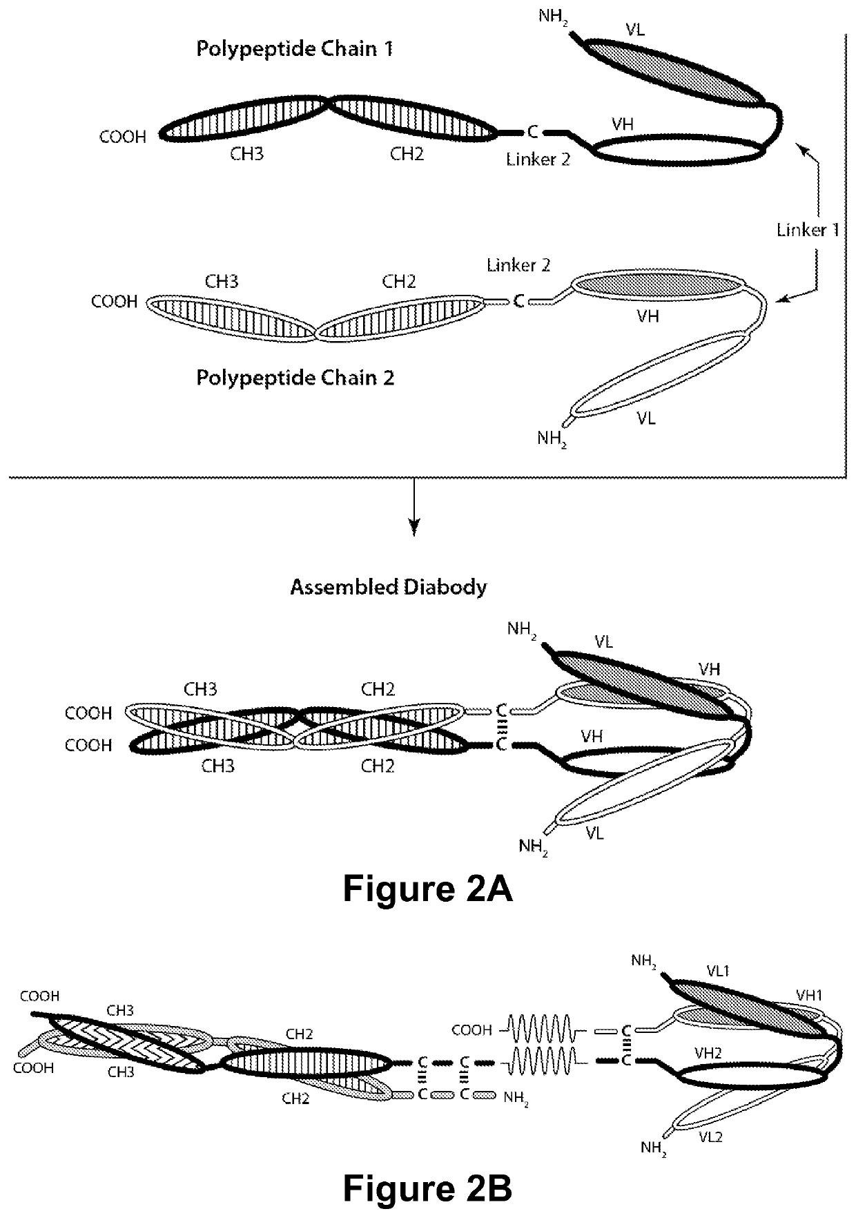
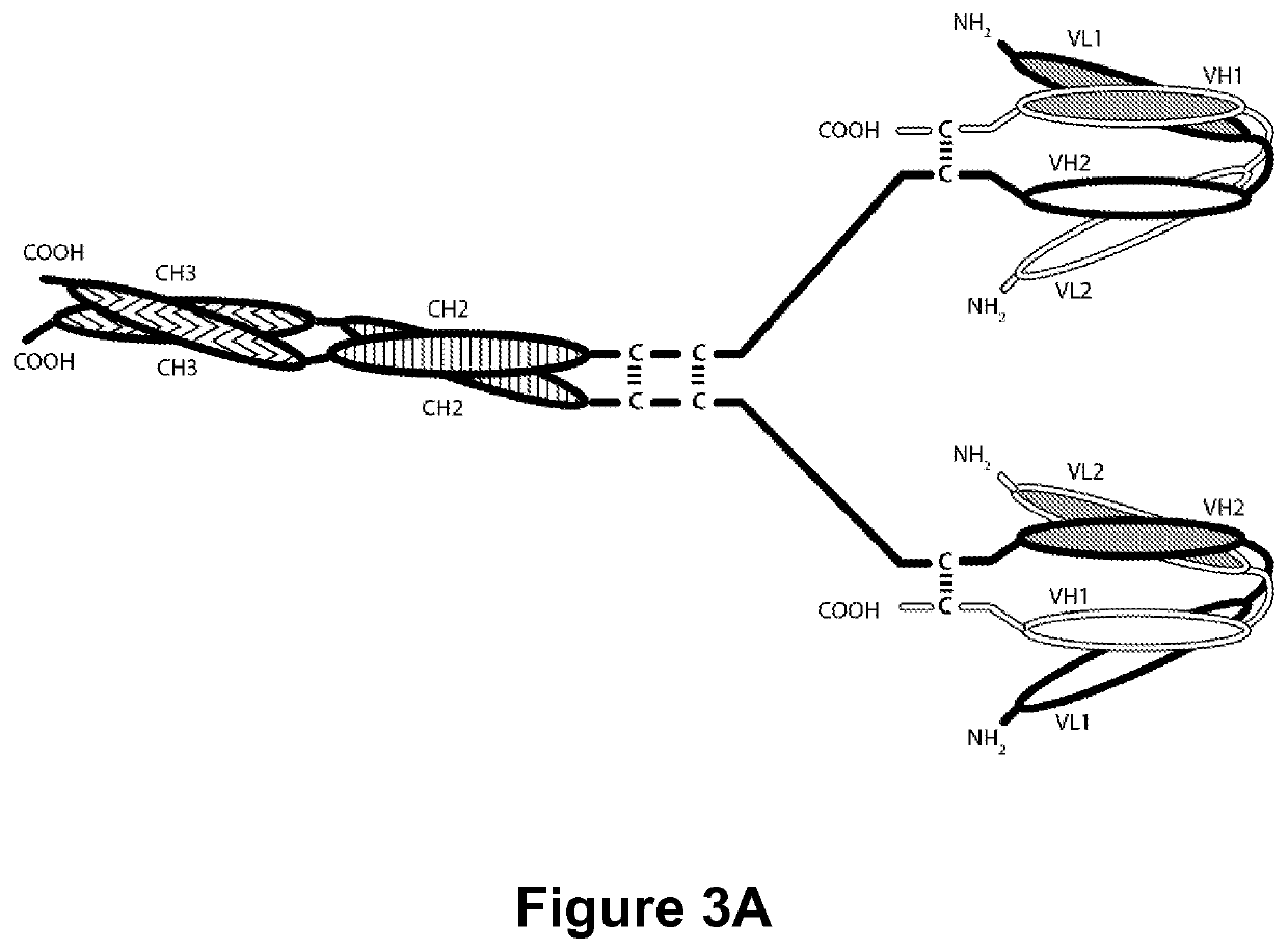
Examples
example 1
W. Characterization of Anti-Human DR5 Monoclonal Antibodies DR5 mAb 1 and DR5 mAb 2
[0940]Two monoclonal antibodies were isolated as being capable of immunospecifically binding to human DR5, and accorded the designations “DR5 mAb 1” and “DR5 mAb 2”. As discussed above, the CDRs of these antibodies were found to differ. In order to determine whether the antibodies bound to different DR5 epitopes, a human DR5-Fc fusion protein was prepared and was coated to an immobilized surface. DR5 mAb 1 (1 μg / mL) was biotinylated and incubated with either a control IgG or with DR5 mAb 2 (10 μg / mL), and the ability of the IgG or DR5 mAb 2 antibody to compete for binding (to human DR5-Fc fusion protein) with DR5 mAb 1 was assessed by measuring the amount of immobilized biotinylated antibody. Additionally, the ability of the IgG or DR5 mAb 1 antibody to compete for binding with biotinylated DR5 mAb 2 was assessed. The results of this experiment are shown in Table 4.
[0941]
TABLE 410 μg / mL Competitor mA...
example 3
Y. Unexpected Superiority of DR5 mAb 1 and DR5 mAb 2
[0950]The ability of DR5-binding molecules DR5 mAb 1 and DR5 mAb 2 of the present invention to mediate cytotoxicity was compared with that of the reference anti-DR5 antibodies: DR5 mAb 3 and DR5 mAb 4. In order to make such a comparison, a bi-specific DR5×CD3 diabody containing the VL and VH Domains of these antibodies and the VL and VH Domains of CD3 mAb 2 were prepared. The prepared diabodies were: DR5 mAb 1×CD3 mAb 2; DR5 mAb 2×CD3 mAb 2; DR5 mAb 3×CD3 mAb 2; and DR5 mAb 4×CD3 mAb 2.
[0951]The employed control diabody contained the VL and VH domains of anti-fluorescein antibody 4-4-20 (respectively, SEQ ID NOs:138 and 139) and the VL and VH domains of CD3 mAb 2 (respectively, SEQ ID NOs:102 and 108), and was designated as the anti-fluorescein×anti-CD3 control diabody “4-4-20×CD3 mAb 2.” The diabody was composed of two polypeptide chains. The first polypeptide chain of the diabody had the amino acid sequence (SEQ ID NO:300) (CDRs...
PUM
| Property | Measurement | Unit |
|---|---|---|
| molecular weight | aaaaa | aaaaa |
Abstract
Description
Claims
Application Information