Detection of membrane proteins

a membrane protein and protein technology, applied in the field of membrane protein detection, can solve the problems of limiting x-ray analysis, hindering progress, and affecting the study of traditional structural biology approaches, and achieve the effect of reducing receptor aggregation and substantially conserving ligand binding activity

Active Publication Date: 2021-03-23
OXFORD UNIV INNOVATION LTD
View PDF4 Cites 0 Cited by
  • Summary
  • Abstract
  • Description
  • Claims
  • Application Information

AI Technical Summary

Benefits of technology

[0008]The present invention is based, at least in part, on a surprising discovery that the use of phosphate ester detergents to form micellar solutions of membrane proteins affords various advantages in the detection of membrane proteins by mass spectrometry. In particular, it has been found that phosphate ester detergents provide for a significant reduction in receptor aggregation, and also allow ligand binding activity to be substantially preserved. Moreover, phosphate ester detergents have been found to be particularly desirable for use in the detection of GPCRs, and may be used to analyse GPCRs, and their interactions with ligands, in a substantially intact, native-like state.

Problems solved by technology

As a significant biological target in disease and cancer, their study by traditional structural biology approaches, such as X-ray crystallography and nuclear magnetic resonance, has been frustrated by limitations relating to their expression and solubility.
Furthermore, X-ray analysis, in the majority of cases, has been limited by crystallographic resolution hindering the assignment of bound moieties.
However, problems of instability and low expression yields have largely hindered progress and GPCRs still remain one of the most important challenges in pharmaceutical research.

Method used


Image

  • Detection of membrane proteins
  • Detection of membrane proteins
  • Detection of membrane proteins

Examples


example 1

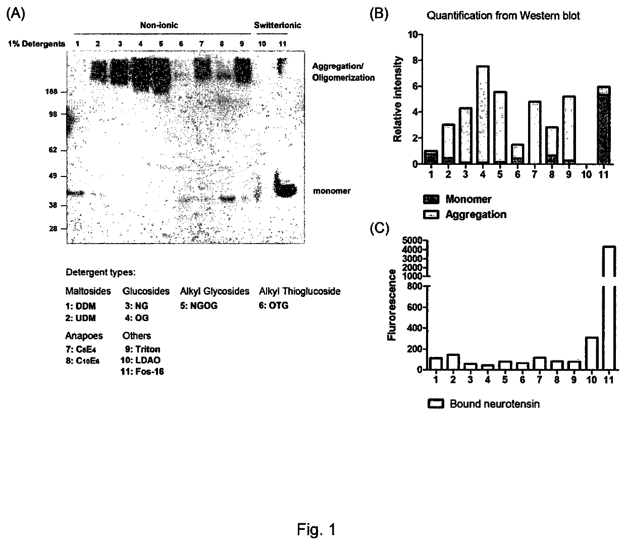
[0138]Various detergents were assessed for their ability to preserve the conformation and activity of NTR1. The detergents assessed were: (1) n-dodecyl-β-D-maltoside (DDM); (2) n-undecyl-β-D-maltoside (UDM); (3) n-nonyl-β-D-glucopyranoside (NG); (4) n-octyl-β-D-glucopyranoside (OG); (5) octyl glucose neopentyl glycol (OGNG); (6) n-octyl-β-D-thioglucopyranoside (OTG); (7) tetraethylene glycol monooctyl ether (C8E4); (8) hexaethylene glycol monodecyl ether (C10E6); (9) polyethylene glycol tert-octylphenyl ether (Triton X-100); (10) lauryldimethylamine-N-oxide (LDAO); and (11) hexadecylphosphocholine (Fos-Choline-16).

[0139]It can be seen from the results presented in FIG. 1 that the use of a phosphate ester detergent (Fos-Choline-16; detergent 11) is advantageous as compared with the other detergents tested. In more detail, FIGS. 1A and B show that less receptor aggregation and oligomerisation occurred with the phosphate ester detergent than with other detergents. FIG. 10 shows that th...

example 2

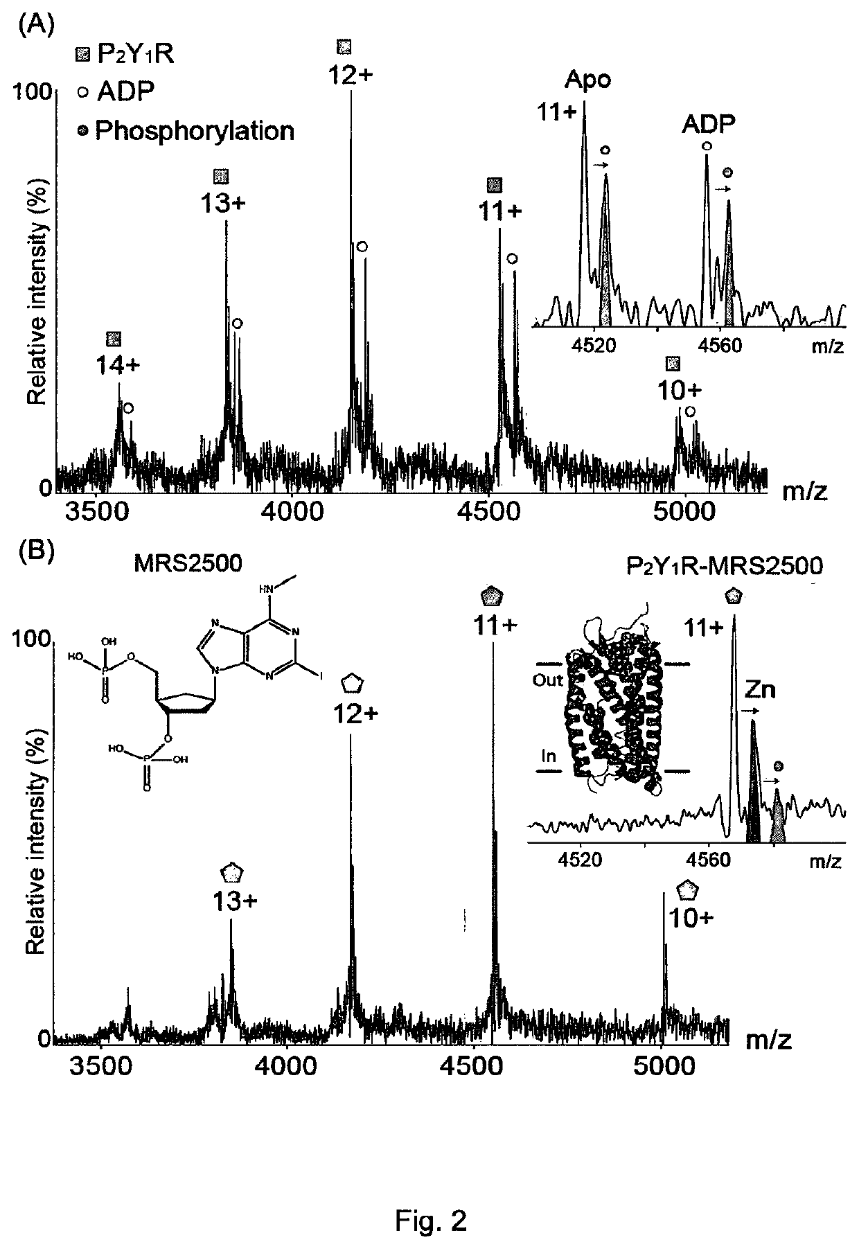
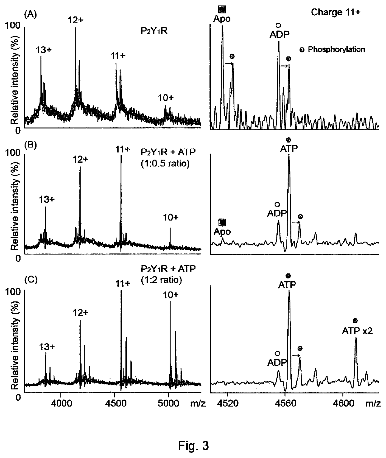
[0140]Mass spectrometry was used to study the human P2Y1 purinergic receptor (P2Y1R) and its interactions with nucleotides and a high affinity antagonist, MRS2500. This receptor, a class A GPCR, functions as a receptor for extracellular adenosine 5′-diphosphate (ADP) and adenosine 5′-triphosphate (ATP), and regulates many physiological processes. P2Y1R is fully activated by ADP to facilitate platelet aggregation via calcium wave propagation and thus serves as an important antithrombotic drug target. Inhibition of P2Y1R activation shows significant decrease in ADP-induced platelet aggregation.

[0141]High-resolution MS was used to probe the drug / ligand interactions of human P2Y1R purified from insect cells. By controlled application of collisional activation, intact P2Y1R was liberated from its detergent micelle, preserving the non-covalent interactions formed in solution. The phosphate-containing detergent of Example 1 (Fos-16) was used in these experiments.

[0142]ESI-MS spectra of pur...

example 3

[0147]In a further experiment, mass spectrometry was used to characterise the binding affinity of PIP2 to NTR1. The phosphate-containing detergent of Example 1 (Fos-16) was used in this experiment.

[0148]FIG. 6A shows the measurement of PIP2 binding of NTR1 in a concentration-dependent manner. Multiple binding of PIP2 was observed after incubation of PIP2 and NTR1 in Fos-Choline-16. FIG. 6B depicts the binding kinetics of various PIP2 to NTR1 (upper; PI(4,5)P2; lower: PI(3,4)P2). Binding curves of different number of PIP2 were plotted according to the quantification of MS spectra, allowing calculation of the dissociation constant (Kd).


PUM

PropertyMeasurementUnit
capillary voltageaaaaaaaaaa
cone voltageaaaaaaaaaa
temperatureaaaaaaaaaa

Abstract

A method of detecting a membrane protein by mass spectrometry comprises: (a) providing a solution comprising a detergent micelle in which said membrane protein is contained; (b) providing a mass spectrometer comprising a nanoelectrospray ionisation source, a mass analyser and a detector; (c) vaporising the solution using the nanoelectrospray ionisation source under conditions such that the membrane protein is released from the micelle; (d) ionising the membrane protein; (e) resolving the ionised membrane protein using the mass analyser; and (f) detecting the resolved membrane protein using the detector; wherein the solution contains a phosphate ester detergent which forms said detergent micelle. Also provided are reagents for use in said method.

Description

FIELD OF THE INVENTION[0001]The present invention relates to the detection of membrane proteins. More particularly, the present invention relates to methods for the detection of membrane protein complexes by mass spectrometry. The methods disclosed herein may be used to detect membrane proteins, including complexes of membrane proteins with ligands such as therapeutic agents.BACKGROUND TO THE INVENTION[0002]Membrane proteins are responsible for a wide range of biological functions. Some of the most prevalent human diseases, including some cancers, result from their dysfunction. Despite representing around a third of the human genome, membrane proteins represent targets for more than half of all current therapeutic agents. As a significant biological target in disease and cancer, their study by traditional structural biology approaches, such as X-ray crystallography and nuclear magnetic resonance, has been frustrated by limitations relating to their expression and solubility. Further...

Claims


Application Information

Patent Timeline
Patent Type & Authority Patents(United States)
IPC IPC(8): H01J49/00G01N33/68C07F9/09H01J49/16
CPCG01N33/6848C07F9/091H01J49/165G01N2333/726G01N2560/00
Inventor ROBINSON, CAROLYEN, HSIN-YUNG
Owner OXFORD UNIV INNOVATION LTD
Why Eureka
Try Eureka