Method and system for plasma assisted low vacuum charged-particle microscopy

a technology of plasma assisted and charged particles, applied in the field of plasma assisted low vacuum charged particle microscopy, can solve the problems of low signal to noise ratio of sem image, inferior resolution and contrast of low vacuum sem and high vacuum sem, so as to reduce beam scattering, improve image quality, and increase detector gain

Active Publication Date: 2021-10-19
FEI CO
View PDF9 Cites 2 Cited by
  • Summary
  • Abstract
  • Description
  • Claims
  • Application Information

AI Technical Summary

Benefits of technology

[0004]In one embodiment, a method for imaging a sample comprises providing a gas in a detection space; providing a magnetic field in the detection space; irradiating the sample with a charged particle beam while providing a plasma in the detection space; detecting charged particles emitted from the sample into the detection space responsive to the irradiation; and forming an image based on the detected charged particles. By providing plasma into the detection space, the gaseous secondary electron detector can perform imaging at lower chamber pressure. Image quality is improved by the increased detector gain and reduced beam scattering.

Problems solved by technology

However, resolution and contrast of the low vacuum SEM is inferior to the high vacuum SEM due to different detector design and increased electron beam scattering in the gaseous environment.
However, the signal to noise ratio of the SEM image remains low due to electron beam scattering in the gaseous environment.

Method used


Image

  • Method and system for plasma assisted low vacuum charged-particle microscopy
  • Method and system for plasma assisted low vacuum charged-particle microscopy
  • Method and system for plasma assisted low vacuum charged-particle microscopy

Examples


Embodiment Construction

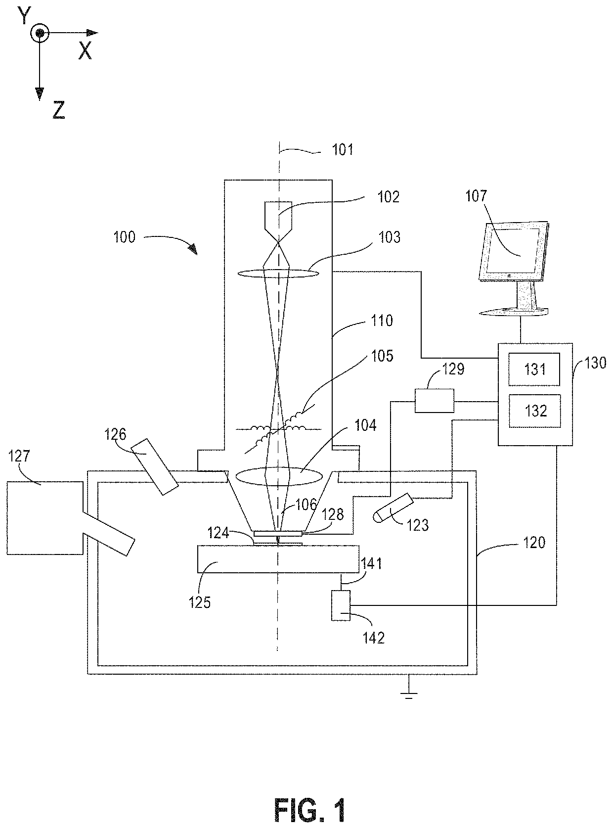
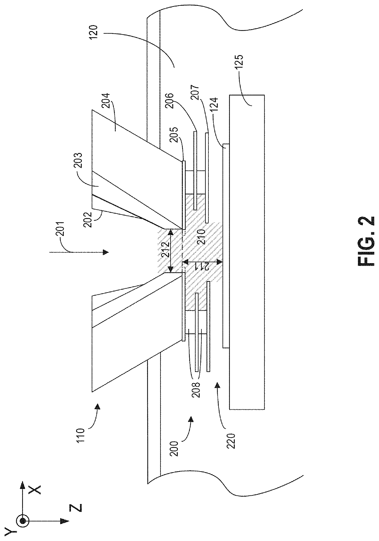
[0017]The following description relates to systems and methods for performing charged particle microscopy in a gaseous environment using a low vacuum microscope, such as a low vacuum scanning electron microscope (SEM) of FIG. 1. The sample positioned in a sample chamber is irradiated with a charged particle beam formed in a particle-optical column coupled to the sample chamber. The sample chamber has a vacuum lower than the vacuum in the particle-optical column. For example, the column, especially the electron source, is pumped to a high vacuum, such as higher than 10−5 torr, for generating the electron beam. The sample chamber may be pumped to 0.01 torr to 50 torr. The difference in vacuum from the sample chamber to the electron source within the column may be achieved via different stages of pumping and multiple pressure limiting apertures (PLAs) positioned along the column.

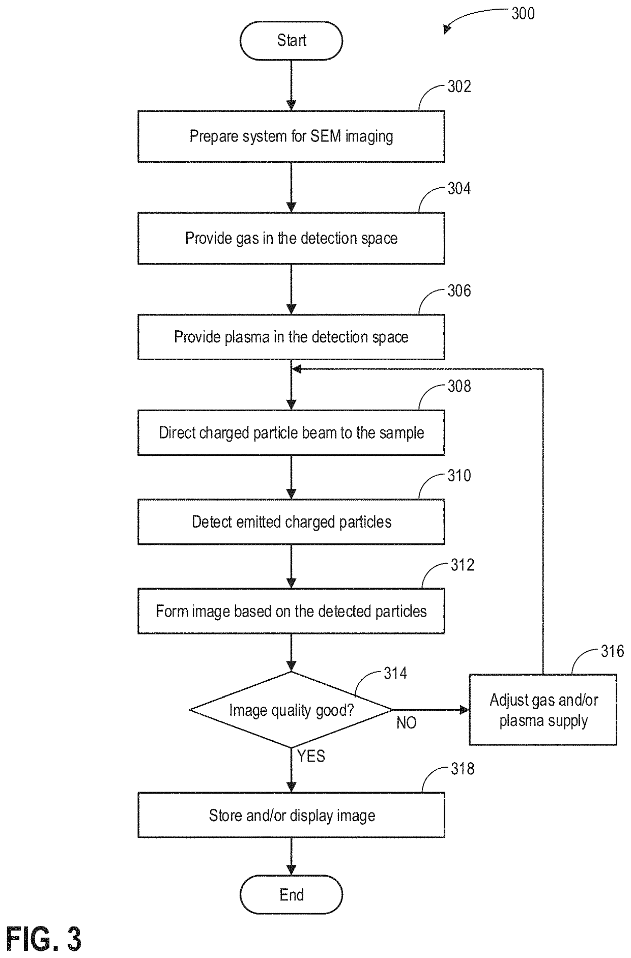
[0018]The low vacuum microscopy system may be operated according to the method of FIG. 3 for plasma assisted...


PUM


Abstract

Various methods and systems are provided for imaging a sample under low vacuum with a charged particle beam. A magnetic field is provided in a detection area of the detector. Gas and plasma are provided in the detection area while detecting charged particles emitted from the sample. Sample image is formed based on the detected charged particles.

Description

FIELD OF THE INVENTION[0001]The present description relates generally to methods and systems for low vacuum charged particle microscopy, and more particularly, to generating high quality sample image based on secondary electrons detected in a gaseous environment.BACKGROUND OF THE INVENTION[0002]Low vacuum scanning electron microscopy (SEM) or environmental SEM allows imaging a nonconductive sample in a gaseous environment using a gaseous secondary electron detector (GSED). Low vacuum SEM provides a means to stabilize sample charging and perform dynamic chemical experiments within an SEM setting. However, resolution and contrast of the low vacuum SEM is inferior to the high vacuum SEM due to different detector design and increased electron beam scattering in the gaseous environment.[0003]One method to improve the image quality in low vacuum SEM is introduced in U.S. Pat. No. 6,972,412. Therein, an electron field and a magnetic field are provided in a detection space above the sample....

Claims


Application Information

Patent Timeline
Patent Type & Authority Patents(United States)
IPC IPC(8): H01J37/244H01J37/28
CPCH01J37/244H01J37/28H01J2237/006H01J2237/2448G01N23/2251H01J37/32449G01N2223/07G01N2223/102H01J2237/2003H01J2237/2449H01J2237/2605
Inventor BISHOP, JAMESTOTONJIAN, DANIELELBADAWI, CHRISLOBO, CHARLENETOTH, MILOS
Owner FEI CO
Why Eureka
Try Eureka