Supercharge Your Innovation With Domain-Expert AI Agents!

Method for imaging a biological sample and corresponding probe

a biological sample and probe technology, applied in the direction of fluorescence/phosphorescence, material analysis using wave/particle radiation, instruments, etc., can solve the problems of difficult detection using tem, osmiophilic and therefore visible for tem, and difficult interpretation of images obtained, etc., to achieve the effect of simplifying correlation

Active Publication Date: 2022-04-12
FOND INST ITAL DI TECH
View PDF1 Cites 0 Cited by
  • Summary
  • Abstract
  • Description
  • Claims
  • Application Information

AI Technical Summary

Benefits of technology

[0039]The osmiophilic precipitate has a high electron-density that allows it to amplify the signal in electron microscopy and therefore make the nanoparticles of platinum clearly visible even at low enlargements and on ample field views.
[0055]In the context of the present invention, the possibility of carrying out simultaneous double localizations of two related molecules in a biological sample means that small probes can be used, since the risk of possible interferences of the probe on the protein of interest is considerably reduced.

Problems solved by technology

Although such probes are able to penetrate inside individual cells possibly present in the biological sample, they are difficult to detect using TEM and require an accretion passage known as silver or gold enhancement.
This accretion passage requires the use of a solution of silver or gold salts in the presence of one or more reducing agents which entail the addition of an aspecific contribution which renders the interpretation of the images obtained problematic.
However, if a double immunolocalization by electron microscopy is to be performed, these probes require the use of complexes consisting of avidin and biotinylated peroxidase which, by bonding with the primary antibody, oxidize the DAB, making it osmiophilic and therefore visible for the TEM.
This is expensive both from the energy point of view and from a cost point of view since it requires long performance times.
This means that the HRP enzyme cannot penetrate inside an individual cell and that it is frequently subject to denaturing in relation to the conditions of the environment in which it is found.
Because of the large sizes compared to the typical sizes of the components of the biological sample, these known lattices can potentially interfere both with the specific molecule associated with them and with the biological sample itself.
Furthermore, these known lattices typically have high levels of toxicity and can therefore significantly alter the behavior of the biological sample.
Some of the known imaging methods for biological samples have numerous disadvantages closely correlated to the size, electron density, and toxicity of the probes used.
Probes with a size in the range of a few nanometers or low electron-density are difficult to detect by TEM, while larger probes can significantly interfere with the antigen-protein activity which does not allow to obtain images without artefacts and / or alterations.
These problems are even more evident in the case where it is intended to detect two different proteins simultaneously, in which pairs of probes of increasing sizes or specific complexes are used.

Method used


Image

  • Method for imaging a biological sample and corresponding probe

Examples


example 1

[0097]To test the actual amplification of the electron-dense signal produced by the peroxidase activity of nanoparticles of platinum in a biological sample inside the cells, in the example shown in FIG. 1 a biological sample containing tumor cell lines (HeLa) was incubated with imaging probes having nanoparticles of platinum of 4 nm and 10 nm.

[0098]The images shown in the squares of FIG. 1 were obtained through electron microscopy, in particular acquired by HAADF (high angular annular dark field detector) scanning TEM (STEM), where the squares “b” and “c” are enlargements of the area 1 highlighted in square “a” and squares “e” and “f” are enlargements of the area 2 highlighted in square “d”.

[0099]With reference to FIG. 1, Applicant has observed, for all the dimensional classes of nanoparticles of platinum used, a considerable increase in the electron-dense signal, which made it possible to identify them even at low enlargement (squares “a” and “d”).

[0100]The images of FIG. 1 acquire...

example 2

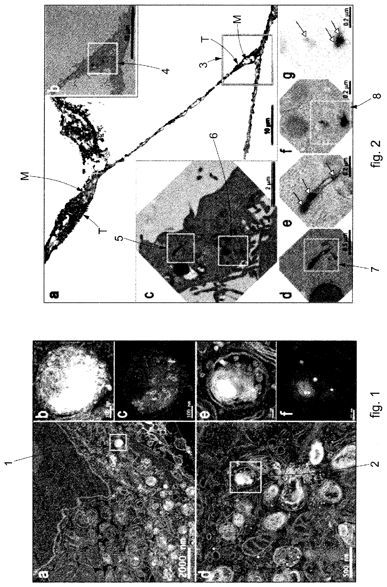
[0103]In the example of FIG. 2, the efficacy was tested of the imaging probes by CLEM to visualize the endosomal pathway of transferrin inside cells.

[0104]In the example shown, the nanoparticles of platinum were bioconjugated with a specific protein, that is, with fluorescent transferrin, suitably functionalizing the surface of the nanoparticles of platinum.

[0105]In particular, this allowed to create an amide bond between the amino groups present on the surface of the protein and the carboxyl groups present on the surface of the nanoparticles of platinum.

[0106]With reference to the square marked by the letter “a” in FIG. 2, the fluorescent signal of the nanoparticles of platinum bioconjugated with fluorescent transferrin is clearly recognizable for the FOM, indicated in black and with the reference “T”, while the mitochondria are indicated in gray and with the reference “M”.

[0107]The localization observed using FOM was confirmed in TEM using the amplification procedure described abo...


PUM

PropertyMeasurementUnit
sizesaaaaaaaaaa
diameteraaaaaaaaaa
sizesaaaaaaaaaa

Abstract

Imaging method for a biological sample using microscopy, for example fluorescence optical microscopy, electron microscopy, or correlative microscopy, which provides to use imaging probes to obtain images in which it is possible to identify the imaging probes and / or possible molecules associated with them. The present invention also concerns the imaging probes, possibly functionalized, that can be used both in CLEM experiments and also in immunocytochemical experiments.

Description

CROSS-REFERENCE TO RELATED PATENT APPLICATIONS[0001]This patent application is a continuation of PCT / IT2018 / 050142, filed Jul. 27, 2018, which claims priority to Italian Application No. 102017000087291, filed Jul. 28, 2017, the entire teachings and disclosure of which are incorporated herein by reference thereto.FIELD OF THE INVENTION[0002]Formulations of the present invention concern a method for imaging a biological sample using imaging probes usable in microscopy, in particular in fluorescence optical microscopy, electron microscopy, or in correlative microscopy (CLEM correlative light electron microscopy).[0003]Here and hereafter, by imaging we mean the acquisition of images of a biological sample or a part of it, such as cells for example, in which it is possible to identify the imaging probes and / or possible molecules associated with them.[0004]The present invention also concerns the imaging probes used to perform experiments on a biological sample by microscopy, such as for e...

Claims


Application Information

Patent Timeline
Patent Type & Authority Patents(United States)
IPC IPC(8): G01N21/64B82Y35/00
CPCG01N21/6486B82Y35/00G01N21/6458G01N2223/418G01N33/533G01N33/54346G01N33/582G01N33/587G01N33/68
Inventor MAROTTA, ROBERTOCATELANI, TIZIANOMOGLIANETTI, MAURODE LUCA, ELISAPOMPA, PIER PAOLO
Owner FOND INST ITAL DI TECH
Features
  • R&D
  • Intellectual Property
  • Life Sciences
  • Materials
  • Tech Scout
  • Unparalleled Data Quality
  • Higher Quality Content
  • 60% Fewer Hallucinations
Learn More