Ultrasound imaging of breast tissue using ultrasound contrast agent

a technology of contrast agent and ultrasound, which is applied in the field of ultrasound contrast agent and ultrasonic imaging of breast tissue, can solve the problems of duct system and small lesions within the duct system that are difficult to identify, and may also be missed in the pathological examination of specimens,

Inactive Publication Date: 2003-06-05
ACUEITY HEALTHCARE
View PDF0 Cites 21 Cited by
  • Summary
  • Abstract
  • Description
  • Claims
  • Application Information

AI Technical Summary

Problems solved by technology

Extension within the ducts may also be missed on pathological examination of the specimen unless a sample slice is taken exactly at the level of the involved duct.
These limitations using current methods of lumpectomy and examination of the specimen may lead to inadequate surgical treatment of the cancer.
Duct systems and small lesions within the duct system may be difficult to identify due to limitations of resolution of the ultrasound machine.

Method used


Image

  • Ultrasound imaging of breast tissue using ultrasound contrast agent
  • Ultrasound imaging of breast tissue using ultrasound contrast agent

Examples


Embodiment Construction

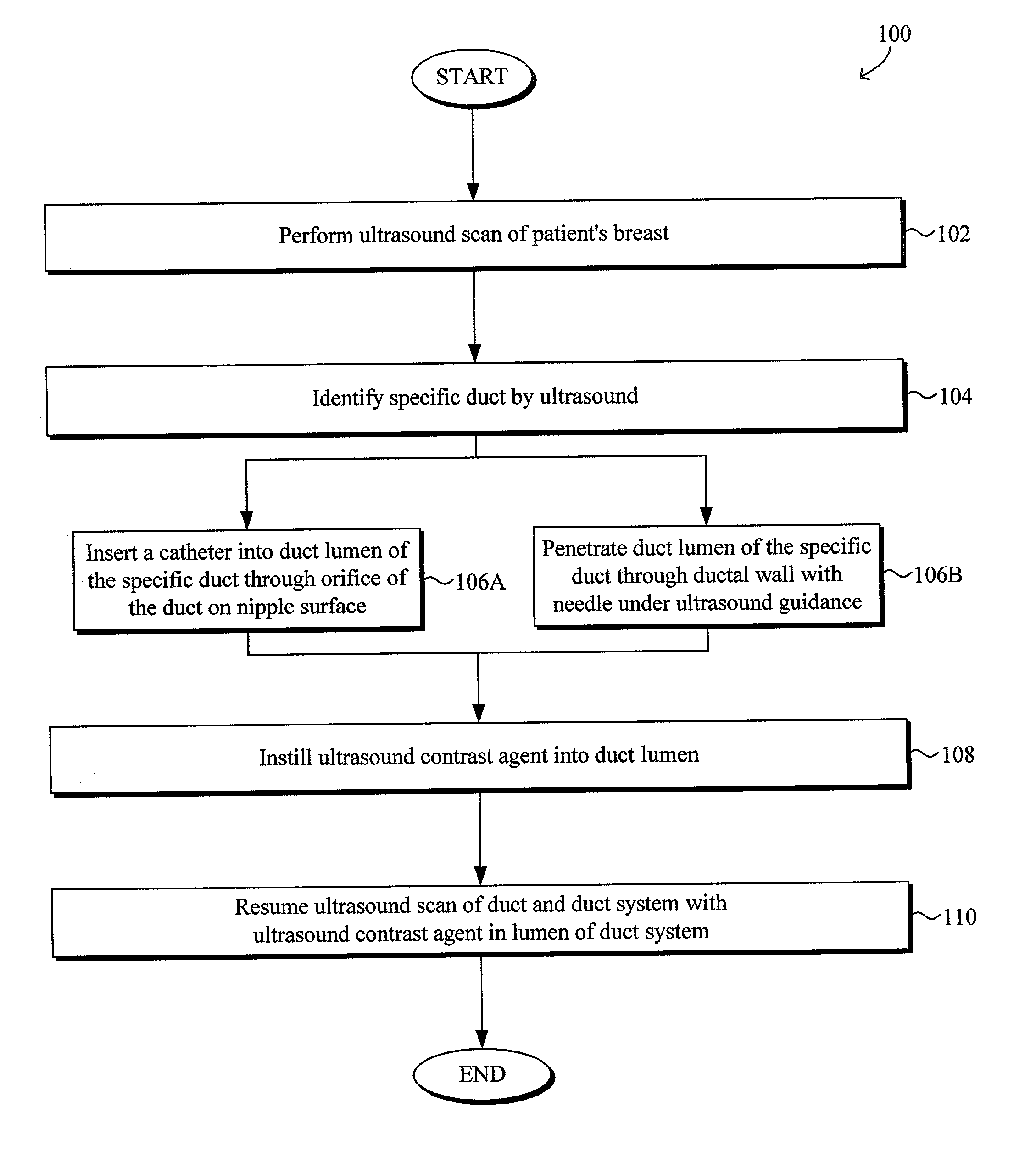
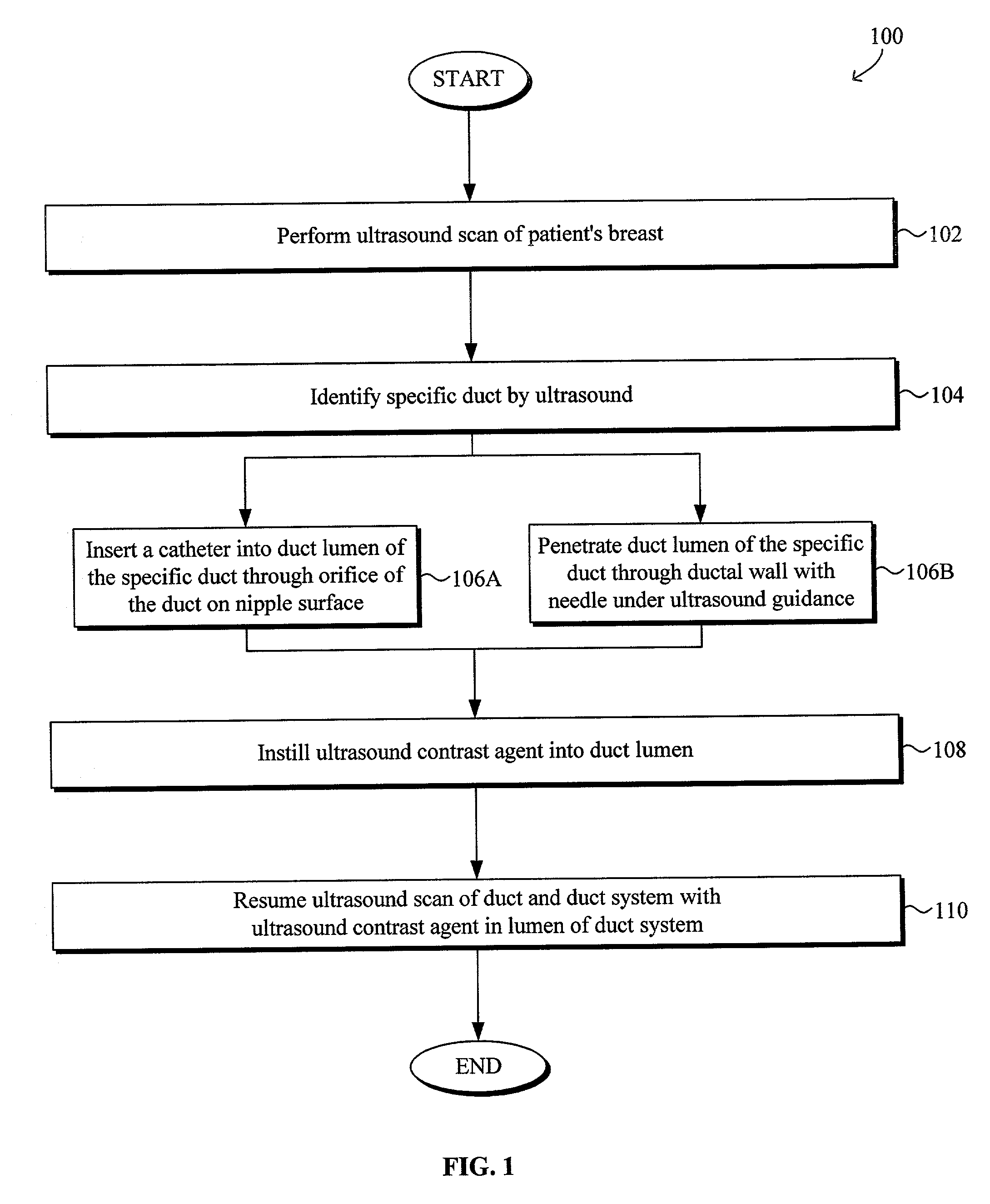
[0014] A system and method for ultrasound imaging of breast tissue by injecting an ultrasound contrast agent into a duct lumen of a patient's breast to enhance the imaging of one or more ducts within a specified lobe of the breast to improve characterization of a lesion or lesions within the duct system of the specified lobe are disclosed. The following description is presented to enable any person skilled in the art to make and use the invention. Descriptions of specific embodiments and applications are provided only as examples and various modifications will be readily apparent to those skilled in the art. The general principles defined herein may be applied to other embodiments and applications without departing from the spirit and scope of the invention. Thus, the present invention is to be accorded the widest scope encompassing numerous alternatives, modifications and equivalents consistent with the principles and features disclosed herein. For purpose of clarity, details relat...


PUM


Abstract

A system and method for ultrasound imaging of breast tissue by injecting an ultrasound contrast agent into a duct lumen of a patient's breast to enhance the imaging of one or more ducts within a specified lobe of the breast to improve characterization of a lesion or lesions within the duct system of the specified lobe are disclosed. The ultrasound contrast agent used to improve breast imaging may be injected into the duct lumen through an orifice on the nipple and/or a duct wall into the duct lumen. The ultrasound contrast agent may be injected prior to and/or during ultrasound imaging. The ultrasound contrast agent may be an acoustically detectable gas such as a halogenated hydrocarbon, halogenated alkane gases, nitrogen, helium, argon and/or xenon. The halogenated alkane gas may be a perfluorinated hydrocarbon such as saturated perfluorocarbon, unsaturated perfluorocarbon, and/or cyclic perfluorocarbon. The acoustically detectable gas may alternatively be mixed in a liquid solution.

Description

[0001] This application claims priority to U.S. Provisional Patent Application Serial No. 60 / 322,634, entitled "Tissue Severing and Removal Devices and Methods," filed on Sep. 12, 2001 and is incorporated by reference in its entirety herein.[0002] 1. Field of the Invention[0003] The present invention relates generally to ultrasound imaging using an ultrasound contrast agent. In particular, a system and method for ultrasound imaging of breast tissue by injecting an ultrasound contrast agent into a duct lumen of a patient's breast to enhance the imaging of one or more ducts within a specified lobe of the breast to improve characterization of a lesion or lesions within the duct system of the specified lobe are disclosed.[0004] 2. Description of Related Art[0005] Surgery plays an important role in the diagnosis and treatment of cancer. In the case of breast cancer, surgery comprises a critical component of medical care where early diagnosis and treatment have demonstrated a significant ...

Claims


Application Information

Patent Timeline
Patent Type & Authority Applications(United States)
IPC IPC(8): A61B8/00A61B8/08A61B17/32A61B18/14
CPCA61B8/0825A61B8/481A61B2018/1467A61B18/14A61B2018/1407A61B17/32056
Inventor LEE, ROBERTA
Owner ACUEITY HEALTHCARE
Why Eureka
Try Eureka