
Method and system for measuring in a dynamic sequence of medical images

a dynamic sequence and medical image technology, applied in image data processing, health-index calculation, sensors, etc., can solve the problems of very limited dynamic information and time-consuming use of the above mentioned method on all images in the image sequen

Inactive Publication Date: 2005-01-27
SECTRA IMTEC
View PDF4 Cites 27 Cited by
  • Summary
  • Abstract
  • Description
  • Claims
  • Application Information

AI Technical Summary

Benefits of technology

[0013] Another object of preferred embodiments of the present invention is to provide an improved method for generating a dynamic sequence of medical images of a moving body part and measuring in said dynamic sequence.
[0015] A further object of preferred embodiments of the present invention is to provide an improved system for measuring in a dynamic sequence of medical images of a moving body part.

Problems solved by technology

Performing the above mentioned steps only once gives very limited dynamic information since only two images in the sequence are used for the measurements.
Thus, using the above mentioned method on all images in an image sequence is rather time-consuming.

Method used


Image

  • Method and system for measuring in a dynamic sequence of medical images
  • Method and system for measuring in a dynamic sequence of medical images
  • Method and system for measuring in a dynamic sequence of medical images

Examples


Embodiment Construction

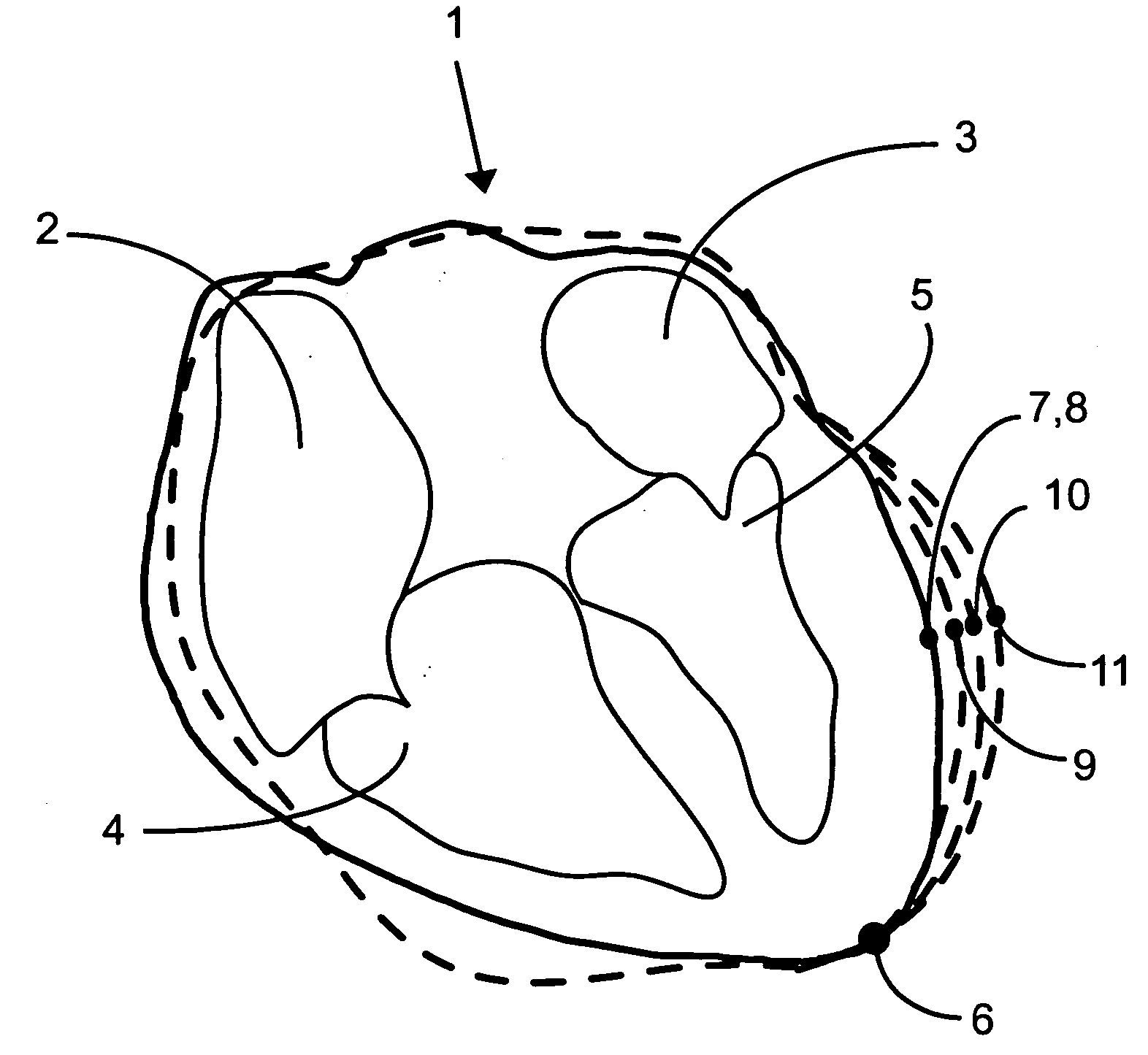
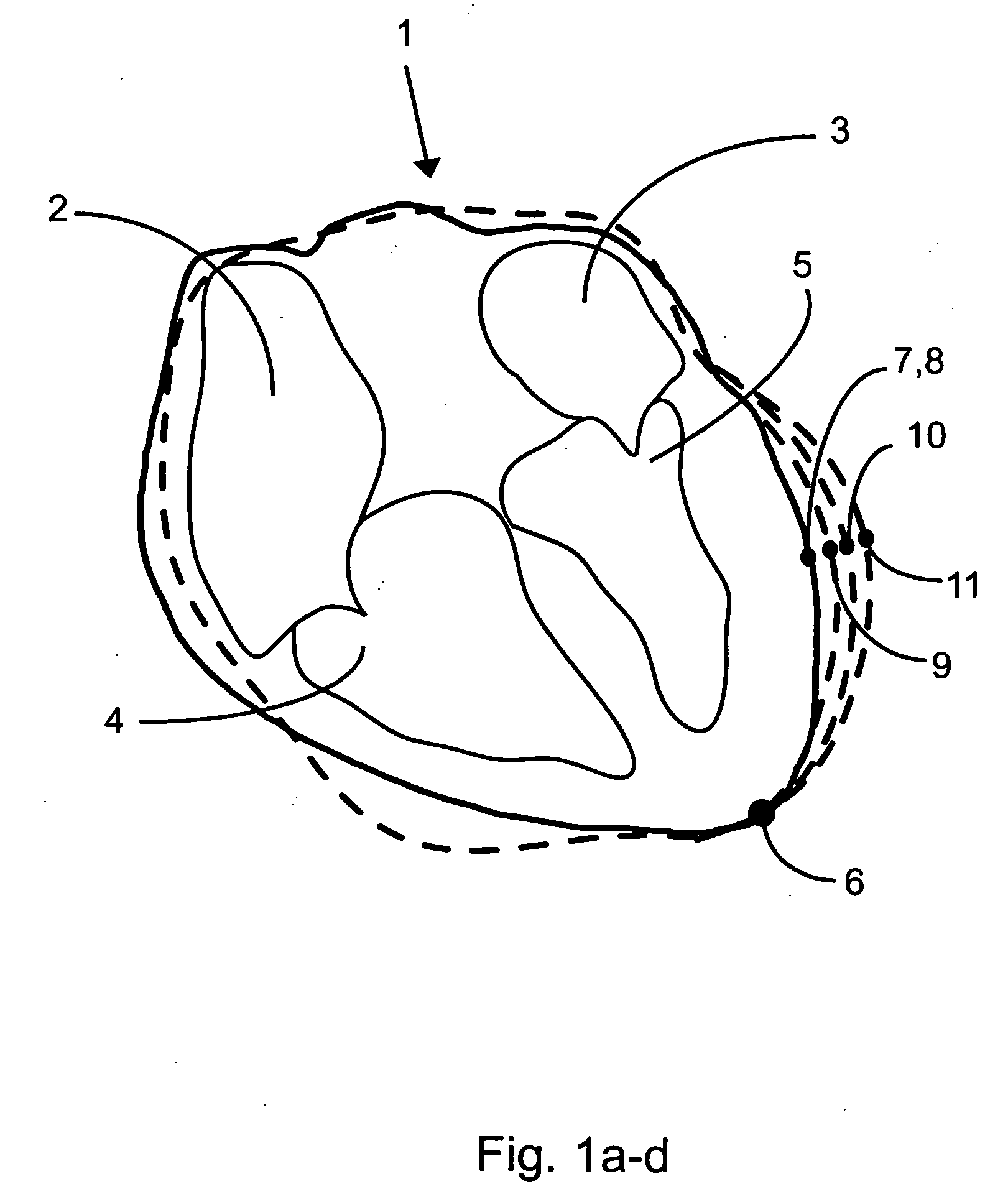
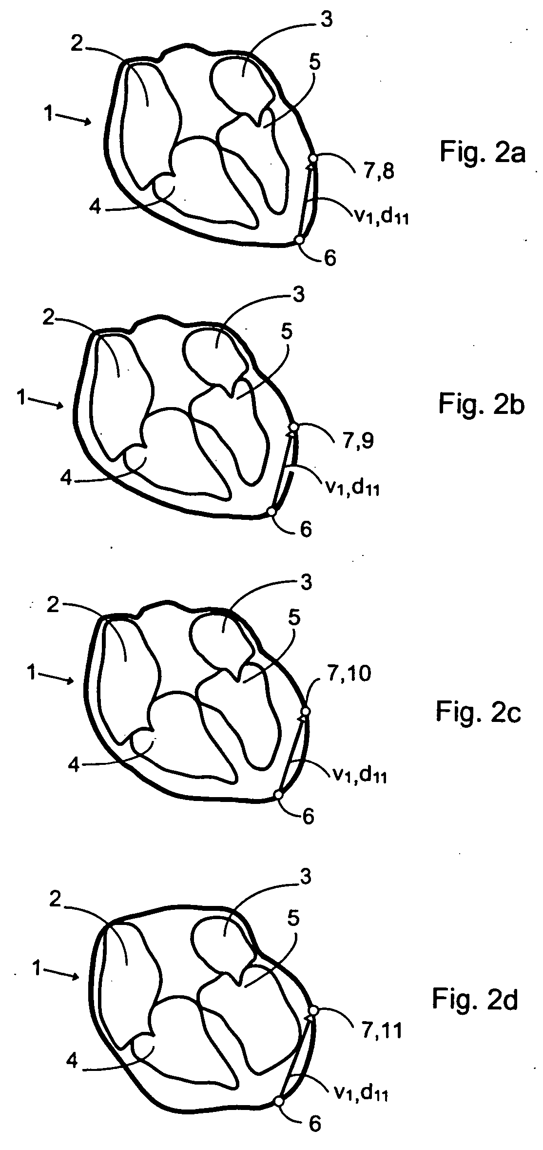
[0031]FIG. 1 shows a composite of highly schematic images of some parts of a heart 1. The images shown in FIG. 1 belong to a dynamic sequence of images of the heart 1, but are only a few images of a dynamic sequence covering a cardiac cycle of the heart 1. The sequence of images may be generated from projection data from time resolved two dimensional X-ray scanning of a portion of a body of a patient including the heart 1. The projection data may thus be time resolved two-dimensional data. In order to cover a complete cardiac cycle the images may be generated at an adequate frequency, for example 12,5 images per second. A contrast medium may be introduced before the scanning to visualize vessels properly on the images.

[0032] The images could be generated at any frequency that permits the analysis of the relevant moving body part. For example, the images could be generated at a rate of from about 9 images per second to about 24 images per second. However, the rate could be greater t...


PUM


Abstract

The present invention relates to a method and a system for measuring in a dynamic sequence of medical images of a moving body part. A reference point being fixed relative to an image geometry and at least one measurement point are defined in the moving body part in one image in the sequence of images. The reference point is then automatically indicated and the at least one measurement point is then automatically tracked in all of the images of the sequence. A length and a direction of at least one vector extending from the reference point to one of the at least one measurement points for each pair of reference point and one measurement point is automatically determined in all of the images of the sequence and at least one of a rate of change of the length and the direction of the at least one vector is automatically determined between selected images in the sequence of images.

Description

TECHNICAL FIELD [0001] The present invention relates to a method and a system for measuring in a dynamic sequence of medical images of a moving body part. BACKGROUND OF THE INVENTION [0002] Non-invasive methods for measuring functions and dysfunctions of body organs typically involve scanning with an X-ray device to obtain projection data for generation of a series of images. When measurements are to be performed of a moving body part, images of several events of the movement have to be created. Images are then generated with a certain frequency, whereby a sequence of consecutive images is formed. The sequence depicts the same anatomical area over a period of time during which some dynamic event takes place and is thus a dynamic sequence. An example of such a dynamic sequence is an image sequence of the heart over one or more heartbeat cycles. [0003] In order to be able to perform measurements in the images, different kinds of measurement tools for application to the images are need...

Claims


Application Information

Patent Timeline
IPC IPC(8): A61B5/107A61B6/00A61B6/03G06T7/20
CPCA61B5/1076A61B6/481A61B6/486G06T2207/30048A61B6/504A61B6/5217G06T7/2033A61B6/503G06T7/246G16H50/30
Inventor YNGVESSON, JONASKAHARI, ANDERS
Owner SECTRA IMTEC
Learn More