

Radiation tomography apparatus
a tomography apparatus and axial scanning technology, applied in the field of tomography apparatus, can solve the problems of generating artifacts, and many scattered radiations on imaging objects, and preventing the degradation of tomographic image contrast, reducing the contrast of tomographic images, and efficiently using.
- Summary
- Abstract
- Description
- Claims
- Application Information
AI Technical Summary
Benefits of technology
Problems solved by technology
Method used
Image

Examples
Embodiment Construction
[0030] Embodiments of the present invention will be described in further detail with reference to the accompanying drawings.
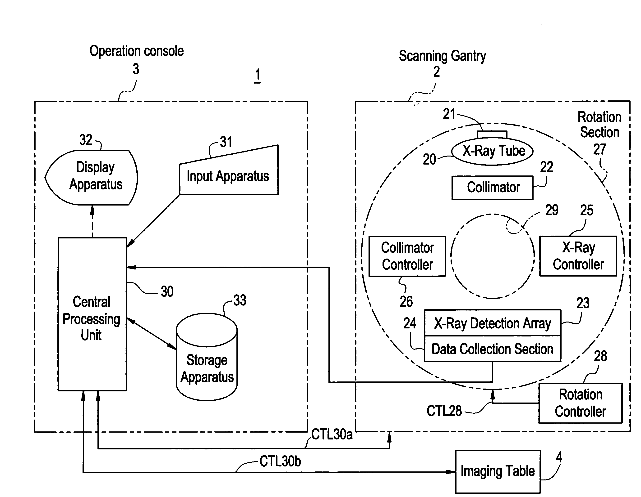
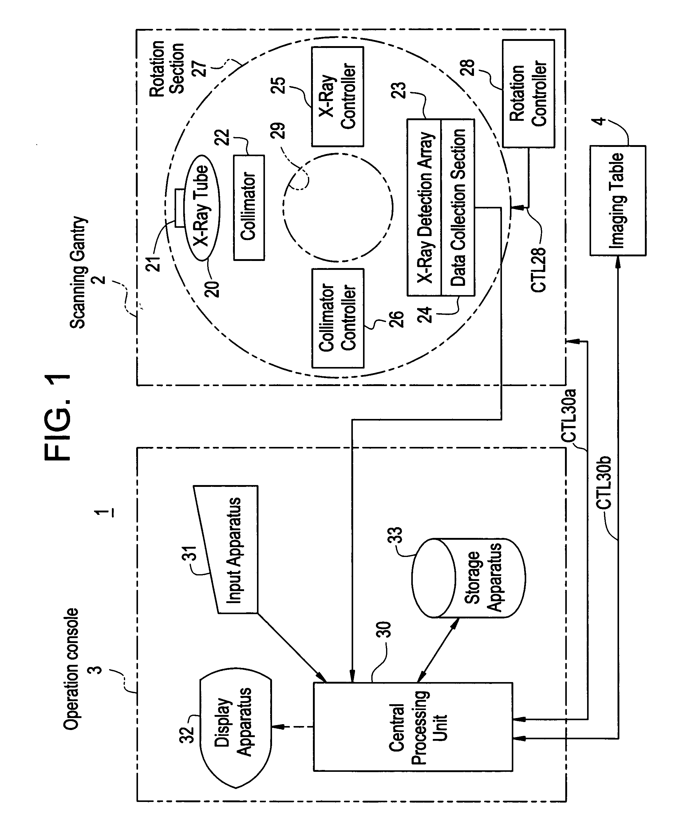
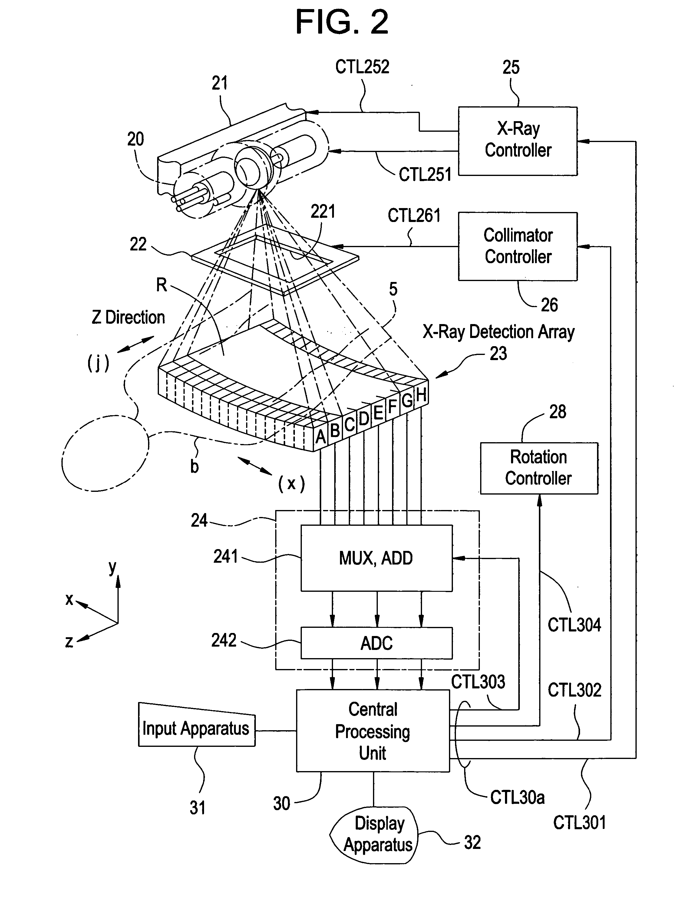
[0031]FIG. 1 is a block diagram showing an overall configuration of an X-ray CT apparatus 1 as a radiation tomography apparatus according to an embodiment of the present invention. FIG. 2 schematically shows major parts of the X-ray CT apparatus 1 as a radiation tomography apparatus according to the embodiment of the present invention.
[0032] As shown in FIG. 1, the X-ray CT apparatus 1 according to the embodiment comprises a scanning gantry 2, an operation console 3, and an imaging table 4.
[0033] The scanning gantry 2 mainly comprises an X-ray tube 20, an X-ray tube moving section 21, a collimator 22, an X-ray detection array 23, a data collection section 24, an X-ray controller 25, a collimator controller 26, a rotation section 27, and a rotation controller 28. The X-ray tube 20 provides radiation irradiation means according to the present invention. The co...
PUM
 Abstract
Description
Claims
Application Information